
Mycogone disease and false truffle of mushrooms in cultivation
18 min read
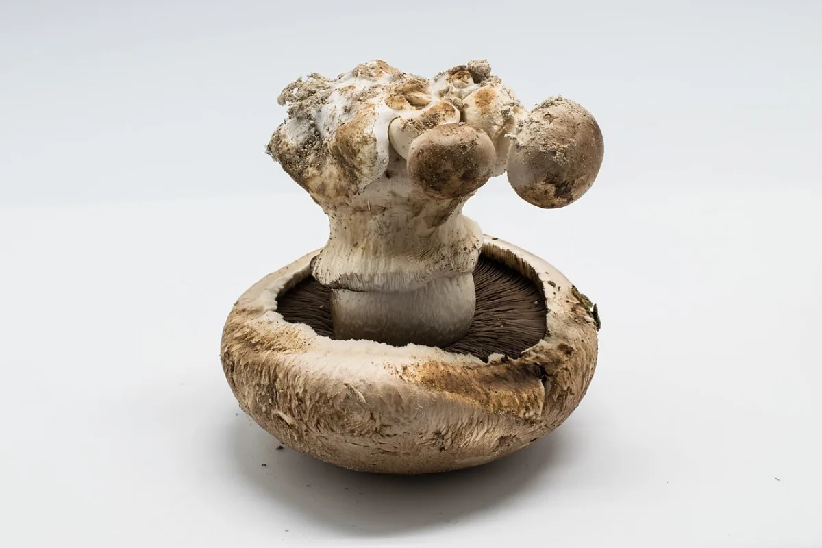
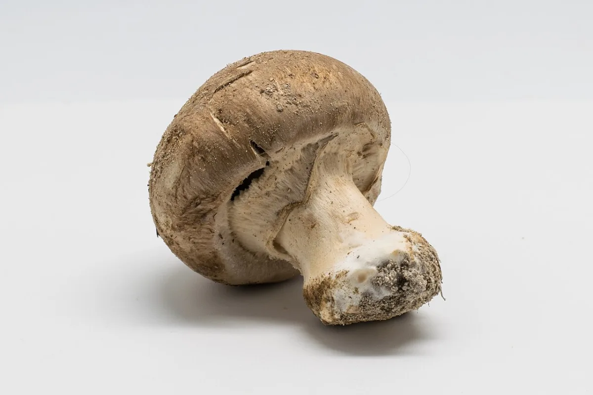
False truffle (Rhizopogon microsporus, syn. Psedobalsamia microspora) p. 266 Red mold, or red geotrichum (Sporen-
Symptoms of this common disease vary depending on the developmental phase of the fruit body during the infection period. If differentiation into a stalk and cap has not yet occurred, a rounded, often large structure is formed, called a sclerodermoid mass. It is often covered with white fluffy mycelium, which gradually darkens and becomes dark brown. Amber-yellow drops of exudate can be seen on the affected tissue. Under the casing soil, the primordia of fruit bodies are sometimes affected; in this case, small clusters of the pathogen's white mycelium appear on the surface of the substrate. Mycogone perniciosa is a pathogen of mushroom fruit bodies that never affects its mycelium.
If infection occurs after differentiation, a brown stripe appears on the stalk, above which a sector of gills colonized by the pathogen can be seen. White mycelium develops on their surface.
Damage to the base of the stalk during the maturation period of the fruit body is accompanied by slight browning without externally visible growth of the pathogen. When left in the substrate, the affected bases of the stalks form abundant aerial mycelium, which gradually turns brown.
.’“ 74 эт и р от « | м д. Ма 2 к - ъж бу г м Г я ь & 4% И феи” 52 *, р 4 р 8 Фо 7}: ЕЛ я иг Г ЧТ Со] 3 Зи № ‹ у = % г. 1 \% УХ. чм. у № +» С > ь 5.2 $ К У, 4 и Ак А а 8 < 42 фа \ к у РР эх > Г Fig. 11.1. Mushrooms affected by Mycogone perniciosa. During severe development of the disease, it is impossible to distinguish the stalk or cap of the mushroom. Such undifferentiated lumps of tissue are called sclerodermoid masses.
M. perniciosa forms two types of spores — thin-walled conidia and aleurospores with a brown thick-walled terminal cell and a thin-walled basal cell. It is assumed that aleurospores remain viable for very long periods, possibly several years.
The period from infection with M. perniciosa to the appearance of symptoms lasts 11 days on average. The pathogen cannot grow through the casing soil; infection occurs only when spores are in close proximity to developing fruit bodies, which apparently stimulate spore germination. The spread of the infection occurs mainly with irrigation water, which, flowing down from the infected beds, carries spores to the healthy crop and simply to the floor of the mushroom house. In addition, workers can be carriers of the infection, as hands, working tools, and boxes become contaminated with spores during crop maintenance. Conidia of M. perniciosa are also spread by air, but this route of spread hardly plays a major role, as does transmission by flies or other insects.
Important reservoirs of M. perniciosa are the soil and plant residues. Hosts of the pathogen include wild mushrooms and some other soil fungi, but their role as sources of infection for the cultivated mushroom has not been studied. In mushroom farms, one of the most common ways of introducing infection into the crop is the use of casing soil contaminated with spores. The appearance of the disease during
рр; ® Г, м. _; 29 р: Fig. 11.4. Sector of affected. г. Г. gills, formed as a result of colonization. 9 и Ления шампиньона Myco- Я ода Ата ви вопе perniciosa at a late ни, м 4 stage of development. the first wave of fruiting indicates that the main source of infection is the casing soil. If white mold develops later, then secondary spread of the pathogen has occurred, most often brought in with working tools or simply by harvesters.
Control. None of the mushroom forms possess resistance to white mold. To prevent the spread of pathogen spores, excessive irrigation and splashing of water should be avoided. Both types of M. perniciosa spores die quickly when heated or treated with a disinfectant solution, although in the latter case — only upon direct contact of the solution with the spores. Therefore, the pathogen often survives under treated surfaces.
If it is suspected that the casing soil is contaminated with spores, it should be subjected to pasteurization (heating to 60 °C for 10 minutes) or treated with a 1% formalin solution, carefully mixing it into the material at a rate of 27 L/m³.
To destroy M. perniciosa, various fungicides are successfully used, for example, benomyl, carbendazim, thiophanate-methyl, and thiabendazole. The preparations are mixed into the casing soil or surface spraying of the beds is carried out. Failures when using benomyl to control white mold in some farms are associated with the biological degradation of this fungicide in the casing soil. Thiabendazole is more resistant to biological degradation. According to recent data, manganese prochloraz is effective against M. perniciosa. In Korea, resistance of the pathogen to benzimidazoles was discovered, but no such reports have been received from other countries.
Verticillium disease (Verticillium fungicola, syn. V. malthousei, V. psalliotae)
This disease can be considered the most common and damaging for the cultivated mushroom. It usually develops particularly strongly in the summer months, when the fly population increases. The disease also flares up in autumn, with high humidity favoring its appearance.
Infection of the developing fruit body before the differentiation into the cap and stalk leads to the formation of structures with undifferentiated tissue ranging from 2—3 to 25 mm in diameter. They are usually grey in color and resemble sclerodermoid masses of white mold, although smaller in size and drier in texture, not as white and fluffy. Infection at a later stage is accompanied by thickening of the stalk, especially at the base. Affected stalks turn brown, the outer layer of tissue peels off and curls downwards. Caps decrease in size, and fine warts appear on them. The affected mushroom often dries out and cracks; as the fruit body grows, the cap deforms and leans downward. Infection of the cap can occur at later stages of crop development. A few days after infection, circular spots with a diameter of
= к а г. № л/а > к: в а м, 2 у 2 # „ МТУ и 2 а Ру Fig. 11.5. Peeling of stalk tissues when infected with Verticillium fungicola.
2» \ я Аб >. р ь, | о а + — Г \ 1.}! Е ы: ^. р { 5 - 54 “ % др 9: а ы аи’ + « % - 4 г й 24 едва я: — и 9 “ “4”; - ле Fig. 11.6. Various deformations of fruit bodies when infected with Verticillium fungicola: a — undifferentiated tissue similar to a sclerodermoid mass during M. perniciosa infection; b — deformation of the cap and stalk, browning of the cap; c — small wart-like outgrowths on a normal mushroom. *: пе а р я. а 3% оао № < ре» щ` ь 40 в ‚_ а зы Е. 1 и. х а р * р в р + \ „ |. Е. Г: — 2 В и, 1 выс ТЫ # = 6; я ыы ьа, # (и 6. в т тай я #. | ИУ * ”: * я И 4 тн. ы р. ий у р} 7 р р а | ож `.. г. Fig. 11.7. Curvature of the cap and browning of the stalk when infected with Verticillium fungicola. ге. к ОР ь № 2”: бы мы. Ач. ть у $. АУ а вы У Fig. 11.8. Brown, light-brown, and sometimes light-yellow spotting of the cap when infected with Verticillium fungicola. about 10 mm appear, initially light-brown, then grey. Sometimes a yellow-pink border is visible around the spots.
Verticillium fungicola infects the mushroom most frequently. Both types of pathogens form numerous conidia; no other types of spores have been detected.
Recently, two subspecies of V. fungicola were identified in the Netherlands. The first, V. fungicola var. fungicola, develops well at a temperature of 24 °C and produces the symptoms on Agaricus bisporus described above. The second subspecies, V. fungicola var. aleophilum, is morphologically very similar to the first, but requires a temperature of 27 °C for optimal growth. On an affected mushroom, it causes brown spotting, without leading to fruit deformation. Due to the higher optimal temperature for development, it is more often found on the bitorquis crop (usually grown at 20 °C, as opposed to the button mushroom, which requires 16 °C) and on the button mushroom in warmer climate conditions.
V. fungicola var. aleophilum has an unusual reaction to benomyl. The ED of benomyl for this subspecies is less than 1 mg/l, but it can grow even at fungicide concentrations above 100 mg/l. For this reason, benzimidazoles in the fight against V. fungicola are morphologically different from V. fungicola.
This subspecies was first discovered on the button mushroom, although it is quite rare here as well. According to the latest data, this subspecies causes brown spotting on bitorquis.
From the initial infection to the appearance of deformation symptoms, it takes an average of 14 days. It is assumed that the germination of the pathogen's spores is stimulated by the mycelium or developing mushroom fruit bodies. The pathogen does not infect the mycelium, but apparently can grow along mycelial strands.
The fungus conidia spread over very short distances, mainly with irrigation water and its splashes. Conidia are formed in sticky clusters and easily adhere to any surface they come into contact with. As a result, they are carried by flies and mites; conidia stick to the hands of workers, to tools, and to harvesting crates. In addition, conidia can be spread by air currents. The dispersal of conidia over a long distance occurs if they are spread by wind or insects, or if they stick to clothing. Both verticillium disease agents are common soil-borne fungi; in the soil, they infect not only Agaricus species but also other fungi.
Control. The most important method of controlling dry bubble is the strict adherence to cleanliness and hygiene in the mushroom house. It is necessary to eliminate flies and ensure that no plant debris enters the facility. Airborne dust, plant debris, and substrate serve as potential reservoirs for both dry bubble pathogens.
Effective control of dry bubble is provided by benzimidazoles; however, currently, due to the widespread use of benomyl, cases of dry bubble caused by benomyl-resistant strains are becoming more frequent. When such strains appear, other fungicides should be used, for example, chlorothalonil, zineb, mancozeb, or the recently approved prochloraz-manganese.
Cobweb disease (Hypomyces rosellus, conidial stage Cladobotryum dendroides, syn. Dactylium dendroides)
Cobweb disease is quite common, although in terms of damage, it is significantly inferior to dry bubble. Affected mushrooms are most often detected during the fourth and fifth waves of fruiting. A typical symptom of cobweb disease is white mycelium, which densely covers the entire fruit body and, in addition, colonizes a significant part of the adjacent casing material. Clusters of mushrooms can be completely covered with cobweb-like mycelium, from which the name of the disease originates. Mushrooms are susceptible to the pathogen at any phase of development. The white cobweb of mycelium turns pink or red with age, and infected fruit bodies become brown and rot.
The pathogen produces numerous spores that are easily spread by air, water splashes, or dripping irrigation water. An outbreak of the disease can occur if the casing material is contaminated with spores carried from an affected crop, although symptoms of cobweb disease are only detected at the last harvests. The pathogen is apparently soil-borne.
Control. Strict hygiene prevents the spread of epiphytotics. Fungal spores die quickly when heated or under the influence of disinfectants. Benzimidazoles are effective against the pathogen, and resistance to them has not yet been detected. However, if mushrooms are simultaneously affected by dry bubble, the use of benomyl against the cobweb disease pathogen can cause an increase in dry bubble. In such a situation, preference should be given to thiabendazole or prochloraz-manganese.
This disease was identified recently and is not widespread. Its most typical symptom is the peeling of mushroom stems, which look shaggy. The color of the stems and caps usually changes, and they gradually become dark brown. A coarse gray mycelium develops on the stems, and it can also be found on the gills of the caps. Sometimes the mycelium colonizes the casing material, which resembles a cobweb disease infection, although the mycelium tissue is thinner. Some affected fruit bodies become stunted and acquire an uneven shape; in other cases (apparently with later infection), only the color of the stems changes slightly, and brown spots surrounded by a yellow border form on the surface of the caps.
Control. The pathogen is a soil-borne fungus, therefore contamination of the casing material or compost with soil and
9 order No. 484 257 m K > +4 7 a, | g No. RU > \ 7 G, v;
E r. mi re |} Sh. =.” m 3 \ (» | r Ch | y + y No. |. —4 m _ ZORE {| 8+ M r. | » ” a | a r \ |, y | | |. k | \ o yyy Ch No. Er -\ y — in m r # “., 4 r: $ $ y - No. 4 „+7 Ya 4 G y r? % i r. ie: A d = r No.“ om Fig. 11.10. Shaggy stipe (pathogen Mycogone perniciosa). Coarse gray mycelium of the pathogen (a) spreads along the stems and gills, affected stems begin to peel (b); some mushrooms are completely covered with a layer of mycelium (c). plant debris can lead to outbreaks of the disease. Of the available fungicides, zineb is the most effective. Cephalosporium spot (Cephalosporium sp.)
Cephalosporium spot is rare on mushrooms. Its symptoms are easily confused with non-pathogenic disorders, in which gills are almost or completely absent on the cap (see p. 271). The gills of mushrooms affected by Cephalosporium spot thicken and become covered with a white fungal coating. The role of the pathogen as the true cause of gill damage has not been definitively established. Sometimes the white powdery mildew-like coating is simply the mushroom's own mycelium developing on the gills.
on which dark brown rounded spots have appeared. The number of spots and their size can vary; sometimes they reach 10 mm in diameter. Often, particles of organic matter are found in the center of such spots. Cap spotting has no economic significance. Bacterial, or brown bacterial blotch (Pseudomonas tolaasii) and ginger blotch (Ps. fluorescens)
Bacterial blotch is one of the most common and damaging diseases of the cultivated mushroom. Its symptoms are the yellowing or light ginger-brown coloration of the caps or dark brown spotting. The cap is affected completely or partially. As the fruit body develops, the affected areas dry out and crack, resulting in asymmetrical caps. Elongated lesions sometimes develop on the stems. The pathogen rarely develops on the gills, although there seems to be some connection between bacterial blotch and bacterial rot (see p. 261). The change in color is usually only superficial, rarely affecting the tissues more than 3 mm deep into the cap. Sometimes signs of spotting appear on mushrooms after harvesting, especially if the harvest is stored at fluctuating temperatures, when moisture condensation begins on the surface of the caps. The pathogen of brown blotch is the bacterium P. tolaasii, and the pathogen of ginger blotch is the closely related bacterium P. fluorescens. Both pathogens were isolated from the casing material, in particular, in the presence of mushrooms affected by bacterial blotch. However, signs of the disease appear most frequently if the primordia of the fruit bodies are heavily infected with bacteria. The incidence is very common when water accumulates in or between mushroom clusters.
It is believed that Pseudomonas tolaasii is widespread in natural conditions. Bacterial cells or infected plant debris are spread by air. The pathogen has been detected in peat and limestone used for preparing the casing material. In addition, the bacteria are spread by flies (in particular, sciarids), nematodes, water splashes, as well as workers.
Fig. 11.11. Brown blotch of the caps during Pseudomonas tolaasii infection, especially severe along the edges (a), and splitting of the caps (b). greenhouse. At 18 °C, the disease develops faster than at 16 °C; high temperatures and relative humidity are particularly favorable for it. Under optimal conditions, the pathogen infects the mushroom and causes blotch within a few hours.
Control. Surface moisture of the caps plays a major role in the development of bacterial blotch. Growing conditions under which water evaporates quickly from the casing material and the surface of the mushrooms generally protect the crop from severe infection. Temperature fluctuations, accompanied by periods of high relative humidity of the air, activate disease development. After irrigation, mushroom caps should be dried as quickly as possible. If bacterial blotch is severe, it is necessary to reduce the number of irrigations and maintain relative humidity below 85% by any means.
Various chemical treatments have been tested in the fight against bacterial blotch, but they do not provide stable results. Chlorination of water at each irrigation (on average 150 mg/l of chlo-
Fig. 11.12. Small black depressions on the surface of the mushroom cap, often shiny. The disease is called bacterial pitting, although its pathogen has not yet been identified. suppresses disease development. Usually, a commercial solution of sodium hypochlorite (10% available chlorine) is used for this purpose at a rate of 0.6 l per 360 l of water. Most prepared solutions of this compound lose chlorine quickly after opening the container, so it is more advisable to buy small quantities of the solution that can be used immediately. Bacterial pitting
The pathogen of this disease has not yet been identified, but it is believed to be a bacterium. Dark brown or black depressions of varying depth, often with a shiny inner surface, appear on the caps of infected mushrooms. The number of such lesions is usually small, but even in this case, they reduce the marketability of the harvest. Most often, bacterial pitting develops during the last waves of fruiting, but the infection is not severe. No specific control measures have been developed for the disease.
Bacterial rot (Pseudomonas cichorii, P. agarici)
This disease occurs quite frequently and is associated with outbreaks of bacterial blotch. Before the differentiation into the cap and the stipe, clear external signs of the infection cannot be detected, but after the veil breaks, they become visible on the gills. The gill tissue often turns dark brown, and small droplets of cream-colored exudate appear on it. Brown lesions also appear on the gills of mature mushrooms, accompanied by cream-colored droplets of exudate. The disease spreads to adjacent gills, and gradually most of them turn into a dark brown mucous mass. The disease is very poorly studied; it rarely reaches the level of an epiphytotic. Corresponding to its nature, the pathogen spreads with water splashes and insects. When the cream-colored exudate dries, the infection can also be spread by air currents.
Control. The same control measures used against bacterial blotch should be effective against bacterial rot, but this has not yet been experimentally proven. Mummy disease (Pseudomonas sp.)
Read next
Plant protection For agronomists
Mummy disease and viral diseases of mushrooms: diagnosis and prevention
Plant protection For agronomists
Biology of fungal pathogens and classification of greenhouse crop disease agents
Plant protection For agronomists