
Mummy disease and viral diseases of mushrooms: diagnosis and prevention
15 min read
Mummy disease of mushrooms: symptoms and protection methods
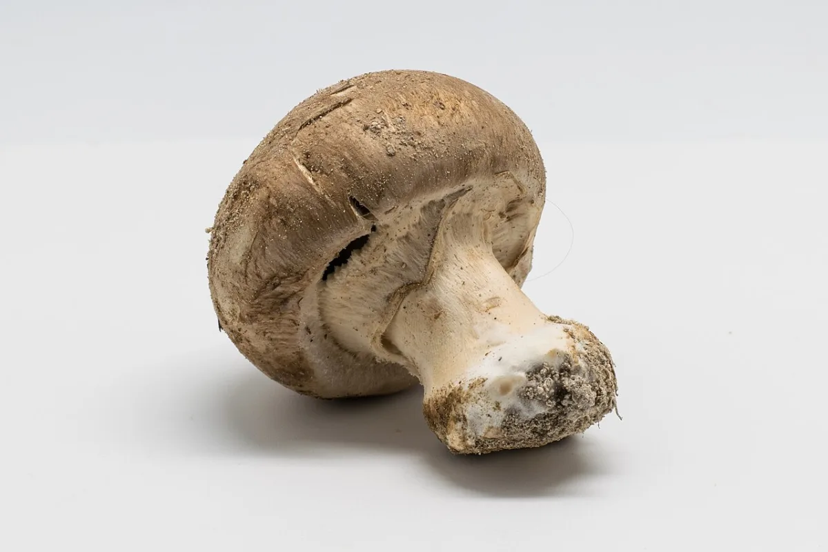
The main sign of mummy disease that an agronomist may notice when harvesting fruit bodies is the strong roughness of the stems to the touch. The disease spreads across the bed or from container to container only through direct contact between healthy and affected mycelium. Because of this, diseased areas often alternate with healthy ones. If the growing conditions prevent the mycelium from spreading to the adjacent bed, this pattern persists throughout the entire growing season.
Outbreaks of mummy disease are closely associated with bacteria of the genus Pseudomonas. In infection experiments, when this bacterium was introduced into the substrate along with the mushroom mycelium, symptoms of the disease appeared in a small number of plots after 2–3 weeks. Other attempts to induce infection using the same organism were unsuccessful.
There are no effective chemical control methods for mummy disease. Reports regarding the increased susceptibility of certain mushroom cultivars to the disease have not been experimentally confirmed.
To contain the spread of mummy disease in the cultivation facility, it is necessary to isolate infection foci in a timely manner:
- Prevent contact between mycelium in different containers.
- On long beds, use the standard barrier method with polyethylene film.
Viral pathologies of mushrooms: diagnostics and sources of infection
Mushroom diseases presumably caused by viruses are known as La France, X-disease, die-back, watery stipe, and browning. The reduction in harvest during their development can be insignificant, but in extreme cases, the crop dies completely. It is important for an agronomist to recognize the specific symptoms of this group of diseases in time.
Typical manifestations of viral pathologies include:
- stem elongation and reduction in cap size (clubbing);
- cap distortion ("German helmet");
- wateriness of the fruit body;
- progressive death of the mycelium, leading to the appearance of gaps in the sowing;
- dwarfing of fruit bodies due to premature ripening.
Direct evidence of the viral nature of these phenomena is absent, but virus-like particles (VLP) are often found in the affected mushrooms. Sometimes VLPs are also found in low concentrations in healthy crops that produce a good harvest. The only precise method for diagnosing VLPs in tissues is electron-microscopic examination.
- Size of mushroom spores — 5–8 µm
- Diameter of small spherical VLPs — 25 nm
- Incubation period of the bacterium in the substrate — 2–3 weeks
Virus-like particles are classified by shape and size. The table below shows the main types of particles found in mushroom crops.
| Particle type | Shape | Dimensions |
|---|---|---|
| Virus 1 | Spherical | diameter 25 nm |
| Virus 2 | Spherical | diameter 29 nm |
| Virus 3 | Bacilliform | 50x90 nm |
| Virus 4 | Spherical | diameter 35 nm |
| Virus 5 | Spherical | diameter 50 nm |
| VLP (described in France) | Club-shaped | — |
The frequency of occurrence of various VLPs has changed over time. In the early 60s, spherical particles with a diameter of 29 nm (virus 2) were the most common, but now particles with a diameter of 35 nm (virus 4) are encountered more frequently. With the exception of virus 5, all other particle types have been isolated from fungal spores. Spores are one of the main mechanisms for the spread of infection.
An indicator of infection is the growth rate of the crop. Mushrooms grown from pieces of fruit bodies with a high VLP content grow significantly slower than normal.
In the absence of a host, the pathogens of viral diseases die quickly. The main sources of infection are mycelium, spores, and crop residues containing VLPs. The mycelium survives on wooden crates and in small compost particles. The wind carries hyphae throughout the mushroom house; they spread particularly actively during the unloading of old substrate.
In cultivation rooms, a huge number of spores with a diameter of 5–8 µm develop, especially when harvesting mature mushrooms. They are carried over long distances by air currents. Wild-growing mushrooms can also be a potential reservoir for VLPs, although they are not the primary source of infection for production facilities.
Spore germination is stimulated by actively growing mycelium. Upon entering the substrate, the spores quickly germinate, after which the fusion of healthy mycelium hyphae and hyphae from VLP-infected spores occurs, thus transmitting the virus to the crop. The greatest losses occur during early infection of a young crop, whereas infection during or after the first flush of fruiting results in only a slight reduction in harvest.
and Fig. 11.14. Micrograph (scanning electron microscope) of mushroom spores on the surface of a fruit body. At such high magnification, it is visible that the fruit body represents a cluster of mycelial strands with large spaces between them. A latent VLP does not always result in a low-productivity crop. The significance of VLP content in low concentrations in the mycelium or mature fruit bodies has not yet been explained, nor has the mechanism of their gradual concentration increase. Virus-like particles are often contained in mushroom tissues, and it is necessary to investigate the role they play in more detail.
Protection against these diseases is closely linked to strict adherence to phytosanitation, that is, adopting measures that are effective against many other mushroom diseases. The main goal of the control program is to protect the new crop from infection by spores and contamination by plant residues from the previous mushroom crop. It is necessary to prevent the introduction of infection resulting from mycelium overgrowth, which leads to the greatest harvest losses.
To protect cultivation facilities from the introduction of mushroom spores via air during ventilation, the following recommendations must be followed:
- Use air filters, ideally with a pore diameter of 1 µm.
- Take all measures to prevent the recirculation of used air.
- Consider the risk of infection in mushroom houses with a common central corridor from which air is supplied to the rooms.
- Filter used air after ventilation to reduce the risk of further spread of virus-like particles.
All wooden parts of cultivation facilities require thorough cleaning after harvesting each crop to destroy all mycelium residues and spores. In mushroom houses where viral diseases cause severe harvest losses, it is advisable to shorten the harvesting period to 4 weeks and pick the mushrooms before the caps open.
Mushrooms differ in their tolerance to viral diseases, and even when virus-like particles are detected in a crop, the reduction in yield may be insignificant. The whitish form, related to forms grown in various cavities, is characterized by increased tolerance. In addition, it withstands a higher concentration of carbon dioxide compared to the common white form, although at the same time, the whitish form develops severe peeling of the caps even with slight air movement. Brown and cream forms also possess tolerance to viral diseases.
Recently, some farms have switched to the cultivation of Agaricus bitorquis instead of the button mushroom, since A. 670415 appears to be immune to VLP detected in the tissues of A. 6{5rogis, and represents a good alternative in case of severe development of viral diseases. It is possible that bitorquis will prove to be susceptible to those viral diseases that do not affect A. 6{5rogis. Rotating mushroom species on a farm will help keep viral infections at a low level.
Many organisms can influence mushroom development both as direct antagonists and as competitors for nutrients. In any case, this reduces the amount of substrate available to the mushrooms, and consequently, their yield drops. Sometimes the development of such organisms serves as an indicator of a compost composition unfavorable for the crop and helps to identify errors made during its preparation.
False truffle (Diehliomyces microsporus, syn. Pseudobalsamia microspora)
The spread of false truffle often leads to mushroom harvest losses. Initially, it is difficult to identify because the well-developed orange-cream mycelium of D. microsporus develops into a felt-like thickening resembling mushroom stroma. However, it differs sharply in color from the grayish or white mycelium of the host. Fruiting bodies of the pathogen can be found in the compost and casing material; they form most abundantly along the edges of the compost layer, particularly in the zone of its contact with the layer of casing material.
The size of D. microsporus fruiting bodies ranges from 3 to 40 mm; their color is initially bright pink, becoming red and then dark brown as they mature. They have a varied shape and a typical wrinkled surface resembling the surface of a brain (hence the English name for false truffle "calves' brains"). The fruiting bodies, or ascocarps, contain many ascospores and release them upon destruction.
If the compost is infected with D. microsporus mycelium, it becomes dark brown, is often highly saturated with moisture, and contains no mushroom mycelium. Frequent gaps are visible on the affected seeded beds; sometimes mushrooms do not develop at all. When false truffle appears during the first wave of fruiting, the mushroom yield can decrease by more than half. A heavily infected crop emits a chlorine odor, which is considered a diagnostic sign of false truffle.
The source of infection is likely the soil, as in some farms cases of particularly severe infection spread were associated with soil excavation during construction work. It was long believed that D. microsporus spores were difficult to destroy. However, recent studies suggest that they cannot withstand exposure to elevated temperatures, even far below pasteurization temperature (60 °C), and that the survival of this fungus is determined simply by the presence of a very large number of spores, as some of them, especially on crates and in cultivation facilities, do not heat up to a lethal temperature. Germination of ascospores is stimulated by mushroom mycelium, especially at elevated temperatures (30 °C), although they can also germinate at lower temperatures (16 °C). Ascospores persist on crates and tools and most often enter mushroom houses with casing material or compost.
Control. It is necessary to avoid contamination of the compost and casing material with soil, otherwise outbreaks of false truffle may occur. Once spores have entered the mushroom house, it is difficult to suppress them. However, since a temperature of 16 °C or higher is required for their germination, the danger of the fungus spreading can be mitigated by maintaining a lower temperature during the mycelium colonization and mushroom harvesting periods. When changing crops, it is necessary to regularly disinfect crates, cultivation rooms, and all work surfaces. All infected crops should be subjected to heat treatment (at least 70 °C for one hour). Constant disinfection of crates and rooms, heat treatment, and filtration (especially during the process of mycelium colonization) allow for the eradication of the false truffle (see p. 273).
In the fight against false truffle, the application of benomyl to compost at a rate of 160 g of Benlate per ton of compost is helpful.
Red bread mold, or red geotrichum (Sporendonema purpurascens)
This is a common competitor of the mushroom, although it rarely causes large harvest losses. The fungus grows in the compost and casing material, forming a thin white coating of mycelium that gradually turns bright pink, then dark yellow. The first colonies often appear in the crevices of the casing layer. As the fungal colonies age, they become red and powdery, releasing a huge number of spores that are carried by the wind. The causal agent of red bread mold is considered to be an antagonist to the mushroom mycelium.
Cracks in the casing material layer can serve as reservoirs for S. purpurascens. Infections of red bread mold can be avoided by strictly following phytosanitary measures on the farm (see p. 273).
Olive-green mold (Chaetomium olivaceum, C. globosum)
The mycelium of Chaetomium species, initially gray, turns white with age and sometimes becomes felt-like. Mature mycelium is often called white chaetomium (though this may be another species). Shortly after mycelium colonization, fruiting bodies—perithecia—are formed, their color changing from olive-green to brown, and their size does not exceed that of a pinhead. Very often, a large number of perithecia develop on particles of straw in the compost.
Mushroom mycelium does not develop in compost infected with Chaetomium species. Infection is often associated with overheating of the compost during the pasteurization process (above 61 °C). In such compost, high concentrations of ammonium ions accumulate by the time of mushroom mycelium colonization, to which Chaetomium spp. are apparently resistant.
Insufficient ventilation of cultivation rooms during pasteurization, over-moistening, compaction, or incomplete decomposition of the compost during its preparation can lead to an accumulation of excess ammonium. If ammonium remains in the compost by the time the mushroom mycelium is sown, the development of Chaetomium species begins.
Apparently, Chaetomium species have an antagonistic effect on the growing mushroom mycelium, and the degree of yield reduction is proportional to the degree of colonization of the compost by the antagonists.
In order to prevent the accumulation of excess ammonium in the compost, it is necessary to control pasteurization conditions by conducting regular ventilation: 28 air volume changes per hour based on the free volume of the room. Sometimes, infected compost becomes suitable for use after extending the pasteurization period for several days at temperatures below 60 °C with active ventilation throughout the treatment period.
Yellow molds and Vert-de-Gris
Various yellow and yellow-green molds sometimes appear on compost regardless of the presence of mushroom mycelium. Once developed, they enter into a competitive struggle with the mushroom mycelium for nutrients or even cause its death through the action of toxins.
Yellow molds can develop under the casing material layer (felt disease, Chrysosporium luteum) or form rounded colonies in the compost (Confetti, C. luteum). Sometimes they inhabit the entire volume of the compost (Vert-de-Gris, Myceliophthora lutea, Sepedonium sp., Sporotrichum sp.).
These fungi produce a multitude of spores that are easily carried by the air. Harvests are reduced to the greatest extent if spores enter the cultivation room during the colonization of the compost by mycelium. Spore populations are reduced when all phytosanitary measures are followed, although if the infection is constantly present, it may become necessary to filter the air entering the cultivation rooms during mushroom mycelium sowing and colonization. If these mushroom competitors develop actively on a farm, it is very difficult to eradicate them.
White powdery mold (Scopulariopsis fimicola)
Secondary mycoses and molds of mushrooms: symptoms and control during the growing process
In addition to major viruses and mummy disease, mushroom growers often have to deal with weed and competitive fungi. They can significantly weaken the growth of mushroom mycelium (Agaricus bisporus) or cause allergies among personnel. Most such infections signal errors in compost preparation—excessive moisture, insufficient pasteurization, or a disruption in the acidity of the substrate.
The appearance of white powdery mold and Coprinus species most often indicates that the compost has not fully decomposed. In this case, white mold actively develops specifically in an alkaline environment at a pH level of 8.2 and higher.
White powdery mold appears as a dense white coating on the casing material or compost, creating an effect of a surface dusted with flour. In the presence of $. Niisoa, the growth of the mushroom slows down, however, after the first waves of fruiting, this pathogen usually dies off, giving way to the crop. Dogaytugre$ species, on the contrary, do not retard mushroom growth, but form dark gray or black fluffy structures in the moist substrate and release volatile spores that are dangerous to the respiratory tracts of workers. The brown mold Rarshazroga Buzuta also prefers dampness: its large, dense colonies are initially white, but quickly turn brown and become powdery.
Green mold (Tisnoyegma ichae, T. Eoptom) acts as a direct antagonist to the mushroom. The pathogen Tisnoyegma ichae colonizes under-decomposed organic matter, dead young mushroom fruit set, and non-disinfected wooden structures, especially those made of immature wood. Another species — Tishoyeorma koshne — covers the surface of the casing material and the fruiting bodies themselves with a fluffy web, leading to their decay (soft wet rot) or the appearance of dry, cracking red-brown spots.
To prevent the development of green mold, it is critically important to follow sanitary rules and hygiene measures (see page 273). Thoroughly disinfect cultivation chambers and minimize the use of untreated wooden structural elements.
Less dangerous is the brown mold caused by the fungi Pe212a oz{ragodegma, RIcapa ria (in the conidial stage — Ozragodaeerma {erregte and Vogyn$ benelia). Their colonies on the casing material change color from white to light brown, sometimes with the formation of brown apothecia. They have only a minor inhibitory effect on the crop and disappear quickly. Conversely, the ink fungi Coprinus (including C. comaus) often germinate before the first wave of mushrooms. As they mature, their caps break down and turn into black slime.
- Alkalinity for white powdery mold — pH 8.2 and higher
- Length of Dogaytugre$ fungal formations — up to 2 mm
- Appearance of Coprinus species — before the first wave
Coprinus spores are easily carried by air currents. If the compost is reliably protected from them during a specific critical period, a reduction in the mushroom harvest will not occur. Protection against the ink cap is structured according to the following scheme:
- Complete the compost pasteurization process.
- Ensure protection of the substrate against the entry of spores from the air.
- Apply the casing material.
Read next
Plant protection For agronomists
Lettuce mosaic virus: symptoms of the disease and methods of plant protection
Plant protection For agronomists
Methods of viral infection spread and plant health improvement techniques
Plant protection For agronomists