
Methodology of clinical examination and anamnesis collection of livestock animals
21 min read
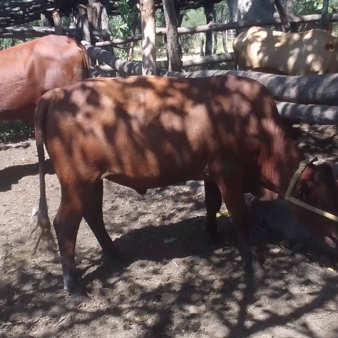
The examination of a livestock animal includes the following stages:
- Registration or recording of the animal in the logbook.
- General examination: habitus, mucous membranes, skin integument, lymph nodes, thermometry.
- Examination of organs and systems: cardiovascular, respiratory, digestive, urogenital, nervous systems, and the musculoskeletal system.
Supplementary examination (bacteriological, virological, serological, blood, urine, feces tests, etc.)
Upon registration, the following data are recorded in the logbook:
| 2 | number or name, which makes it easier to identify the animal in a large farm |
Anamnesis — a set of information obtained about a sick livestock animal through questioning. Life history: where and how it is kept, feeding peculiarities, nature of use, and whether it has been ill before.
Case history: the purpose of the anamnesis is to clarify everything that can provide a correct understanding of the causes, signs, and duration of the disease. While collecting an anamnesis, it is necessary to obtain answers to the following questions:
- When did the animal get sick? At the same time, it is checked whether the animal has been ill before.
- Why and under what circumstances did the disease occur, i.e., what could have been the cause of the disease?
- What signs of the disease were noticed? When collecting this information, questions are asked in a form understandable to the respondent; in particular, when trying to find out how well the digestive system is functioning, one asks about the animal's appetite, what feed it prefers, whether there was bloating or diarrhea, etc.; when clarifying the activity of the respiratory system, one asks whether there was coughing, shortness of breath during work, etc.
- Are there any other sick animals on the farm with the same signs? This question clarifies the farm's status regarding infectious diseases and sometimes helps identify other mass diseases caused by poor feeding, housing, and care.
- Was treatment applied, what kind, when, and by whom?
All the listed questions are not always sufficient to clarify the causes and essence of the disease. Therefore, depending on the conditions, their quantity and nature may change.
Having collected the anamnesis, one proceeds to the general examination of the livestock animal. This examination consists of determining the animal's habitus, examination of visible mucous membranes, lymph nodes, coat, skin, and subcutaneous tissue.
The data obtained during the general examination in some cases allow for identifying the main symptoms of the disease. In addition, through general examination, one can notice a sick livestock animal and separate it from the healthy ones during mass inspections of animals in the pasture and when walking around cattle yards.
Determination of the animal's habitus. Habitus is understood as the state of the animal, which is characterized by the body build, condition, body position, and temperament of the animal.
Body build is usually determined by inspection. At the same time, attention is paid to the development of the skeleton, muscles, skin, subcutaneous tissue, and the ratio in the development of individual parts of the body.
Condition is determined by inspection and palpation. Depending on the volume of musculature and subcutaneous tissue and the roundness of the body contours, good, medium, and unsatisfactory condition are distinguished.
The body position can be normal and forced or abnormal. When determining various abnormal positions, one can establish symptoms of many diseases and notice a sick animal while walking around the herd. For example, in case of pharyngeal inflammation, the animal keeps its head extended; during parturient paresis, a cow lies with its head on its chest, not reacting to irritation.
Temperament is determined by the behavior of the animal. Calm, lively, excitable, and phlegmatic temperaments are distinguished:
- The calm temperament is characterized by usual mobility without increased reaction to the environment.
- With a lively temperament, animals are more mobile and energetic.
- With an excitable temperament, in addition, excessive excitability and nervousness are observed.
- The phlegmatic temperament manifests itself in lazy and sluggish movements and weak reaction to the surroundings.
Evaluation of temperament is important when examining and treating an animal, as animals with lively and excitable temperaments usually react more strongly to pain and are more dangerous to the person examining them or providing them with assistance.
Examination of mucous membranes. During a general examination, one can limit oneself to the inspection of the conjunctiva and the eye membrane. Other mucous membranes (nose, mouth, vagina) are examined when examining the corresponding organs.
When inspecting the eye membranes and eyelids of a horse, the palpebral fissure is opened with the thumb and index finger, while holding the horse's head with the other hand. In cattle, when inspecting the sclera, the head is turned to the side, and the mucous membranes of the eyelids are examined in the same way as in a horse. In small animals, it is more convenient to open the palpebral fissure with both hands.
In a healthy state, the conjunctiva has a pale pink color. In case of diseases, the following changes may be observed in the conjunctiva:
- pallor of the conjunctiva - during blood loss and chronic diseases associated with exhaustion;
- redness (hyperemia) - during many infectious diseases, poisoning, and intoxication;
- jaundice - during liver diseases and certain blood diseases;
- cyanosis, or bluish discoloration - during heart and lung diseases;
- hemorrhages (bleeding) - during purpura and sepsis.
Figure 24 – Examination of the mucous membranes of the eyes and eyelids: A (left) – in a horse,
Examination of lymph nodes and skin
Clinical examination begins with palpation of superficial lymph nodes. In cattle, the mandibular and prescapular nodes, as well as the nodes of the knee fold and udder, are easily palpated. In horses, the mandibular nodes are normally easily palpable. In case of pathological enlargement, it is possible to palpate the parotid, cervical, and inguinal nodes in horses, and in cattle — the upper and middle cervical, as well as retropharyngeal nodes.
During palpation, the specialist determines the size, shape, character of the surface, consistency, temperature, sensitivity, and mobility of each node. These parameters are critical for the timely diagnosis of dangerous infections, such as glanders, strangles, and tuberculosis. Any deviations from the norm require immediate control of the situation.
Changes in the skin and coat reflect not only dermatological problems but also violations of housing and feeding conditions, as well as the development of severe internal diseases.
The condition of the skin is assessed by visual inspection and palpation, if necessary, by additionally examining scrapings under a magnifying glass or microscope. The veterinarian checks the state of the coat, temperature, humidity, elasticity, odor, color, and skin sensitivity. Hair loss and changes in the coat are characteristic of poor care, as well as mange, ringworm, and eczema. Dryness of the nasal mirror in cattle, the snout in pigs, or the nose in dogs signals a disease, and a general decrease in skin temperature often accompanies heavy blood loss or milk fever.
During the examination, it is important to record the presence of itching, edema, nodules, and any breaches in the integrity of the skin. Wounds, scratches, cracks, pressure sores, and gangrenous areas indicate mechanical damage or systemic failures in the animal's body.
Thermometry and diagnostics of the cardiovascular system
Body temperature measurement is carried out with a special veterinary medical thermometer, the mercury column of which records the maximum value until it is shaken down. The procedure requires strict restraint: cattle are held by the horns or with a nose lead, horses have their front leg lifted, pigs are calmed by scratching or laid down, and dogs are restrained by the head.
- Disinfect the thermometer with alcohol or a disinfectant solution and lubricate it with petroleum jelly or oil.
- Stand on the left side of the animal, move the tail aside, and carefully, rotating slightly, insert the instrument into the rectum. In small animals, the thermometer is inserted 1/3–1/2 of its length to prevent rupture of the intestine; in case of diseases of the anus, the vagina is used.
- Hold the thermometer by hand or with a holder in the rectum for at least 10–15 minutes.
- After removal, clean the instrument of feces and mucus with warm water and disinfect it in a vessel with alcohol.
- Normal temperature of cattle — up to 39,5 °C
- Normal temperature of a horse — up to 38,5 °C
- Procedure time — 10–15 minutes
- Insertion in small animals — 1/3–1/2 of the length
Temperature deviations above the norm, not caused by physiological factors, indicate the development of a disease. Recording the indicators by day forms a temperature curve: a continuous increase indicates a worsening of the condition, and a gradual decrease indicates that recovery has begun.
| Type of animal | High temperature threshold |
|---|---|
| Cattle | above 39,5° |
| Horse | above 38,5° |
A persistent and rapid drop in body temperature below the norm (subnormal temperature) is observed in milk fever and acetonemia. Such a state can be a sign of an approaching fatal outcome.
Examination of the cardiovascular system includes auscultation, heart percussion, and assessment of the impact of the discovered pathologies on the entire body. Special attention is paid to the heart impulse — the protrusion of the chest wall during the contraction of the ventricles, which is assessed by inspection and palpation. In healthy animals, the impulse is felt with the palm as a moderate, light vibration. Strengthening of the impulse indicates an increased load on the heart; weakening indicates heart weakness.
| Type of animal | Localization of the heart impulse | Area |
|---|---|---|
| Cattle | Left in the 4th intercostal space | 5–7 sq. cm |
| Horse | Left in the 5th, right in the 4th intercostal space (8 cm below the line of the scapulohumeral joint) | 4–5 sq. cm |
Heart percussion allows for the precise determination of its borders, size, and sensitivity. Expansion of the dullness area or manifestation of pain during percussion serve as important diagnostic signs of pathological processes, for example, inflammation of the pericardium.
Cardiac auscultation. During auscultation, or listening to the heart, the ear is pressed firmly against the area where the heart is located, or a stethoscope or phonendoscope is used. During this procedure, the livestock animal is held by the head, and the thoracic limb on the side being examined is pulled slightly forward. When listening to the heart, the heart sounds, their intensity, and any possible murmurs are determined.
Heart sounds. Heart sounds are heard as two successive sounds. In large animals, both sounds can be roughly represented by the sounds "lub-dup". Fainting of the heart sounds is observed in cases of inflammation of the heart muscle and pericardium.
Heart murmurs. Heart murmurs arise in connection with abnormal heart activity and differ sharply from heart sounds. According to the place of their formation, endocardial (or intracardiac) and extracardiac (or extracardiac) murmurs are distinguished. Endocardial murmurs are most often observed in cases of endocarditis or so-called heart defects and sometimes in diseases of the heart muscle. In their nature, endocardial murmurs resemble the sound of a bumblebee flying, hissing, and whistling.
Examination of the arterial pulse. The pulse is examined by placing a finger on an artery.
In cattle, the external maxillary artery is examined where it runs along the lower edge of the masseter muscle. In addition, the pulse can be felt on the femoral artery, which runs in the middle of the inner surface of the hind limb, and the middle artery of the tail, felt on its inner surface.
In the horse, donkey, and mule, the pulse is usually determined by the external maxillary artery, which runs on the inner surface of the lower jaw.
In small animals, the pulse is most often examined on the femoral artery.
When examining the pulse, the number of pulse beats, its quality, and rhythm are determined. The number of pulse beats is always calculated per one minute of time. Table 2 shows the limit fluctuations of the pulse in healthy animals.
Abnormal changes in the pulse are considered to be acceleration, weakening, and arrhythmia of the pulse. Pulse acceleration (tachycardia) is observed in many heart diseases and some infectious diseases. A weak pulse usually occurs with cardiac weakness and with a decrease in the tension of the vessel walls (their tone). An arrhythmic pulse manifests itself in the form of irregular alternation of pulse beats and unequal magnitude of the pulse wave. It is most often observed in heart disease.
Table 2 – Number of heart contractions (pulse) per minute in healthy animals
Animal species Pulse Animal species Pulse Horse 24-44 Donkey 42-52 Cattle 50-80 Deer 36-48 Pig 60-80 Dog 70-120 Sheep and goat 70-80 Cat 110-120 Camel 32-52 Rabbit 120-140 Mink 90-180 Chicken 120-150 Blue fox 90-130 Duck 140-250 Silver-black fox 80-140 Goose 120-160
Of the additional methods for examining the cardiovascular system, electrocardiography and blood pressure measurement are more often used.
Electrocardiography is the recording of the electrical currents of action and the working heart muscles using a special device called an electrocardiograph. This recording is called an electrocardiogram and often allows for obtaining the most valuable indications for the diagnosis of many pathological conditions of the heart.
An oscillosphygmometer is used to measure blood pressure. This method is used to identify high blood pressure (hypertension) and low blood pressure (hypotension).
Respiratory organs are examined by inspection, palpation, percussion, and auscultation. Possible causes and signs of the disease are determined in advance through anamnesis, and body temperature is measured.
2) to examine the upper respiratory tract and paranasal sinuses;
Examination of respiratory movements. When examining respiratory movements, the following are determined: 1) the number of respiratory movements; 2) the rhythm of breathing ; 3) the strength of respiratory movements.
Number of respiratory movements. The number of respiratory movements is determined by observing:
1) the rise and fall of the chest;
2) the movement of the groins or abdominal muscles;
Respiratory movements are counted for one minute. The number indicated in Table 3 is considered normal.
Table 3 – Number of respiratory movements per minute in normal adult animals
Animal species Number of resp. movements per minute Animal species Number of resp. movements per minute Horse 8-16 Dog 15-30 Cattle 10-30 Cat 20-30 Sheep and goat 12-20 Rabbit 50-60 Pig 10-20 Chicken 12-30 Camel 2-12 Duck 16-30 Reindeer 8-16 Goose 12-20 Mink 40-70 Pigeon 16-40 Silver-black fox 14-30
An increase or decrease in the number of respiratory movements compared to these limits should in most cases be regarded as a sign of disease.
Examination of the upper respiratory tract and paranasal sinuses. The following are subjected to examination:
1) nasal discharge and exhaled air;
Percussion of the chest. Percussion, or tapping, of the chest is performed with a percussion hammer and a pleximeter. During percussion, the pleximeter is pressed firmly against the surface of the intercostal spaces. The blow with the percussion hammer is delivered vertically and quickly two or three times in a row. When determining the boundaries of the lungs, percussion usually begins from the front to the back along horizontal lines drawn from the tuber coxae, the ischial tuberosity, and the scapulohumeral joint. To reveal the condition of the lungs and pleura, percussion is usually performed from top to bottom along the intercostal spaces.
Auscultation of the thorax. Auscultation, or listening to the chest, is used to determine the nature of sounds originating from the activity of the respiratory organs. These sounds are referred to as breath sounds.
Lung auscultation and respiratory organ X-ray diagnostics
Thoracic auscultation is performed by applying the ear directly through a towel or by using a stethoscope and phonendoscope. When listening to the anterior sections, the specialist stands to the side, facing the head of the livestock animal, while keeping a hand on the withers or back for self-protection. The anterior regions of the left side of the thorax are auscultated with the right ear, and the right side with the left ear. To examine the posterior sections of the lung, one should stand facing the rear of the livestock animal.
To protect yourself from being kicked by the hind limbs of restless horses, it is necessary to lift and hold a thoracic limb, as well as securely fix the head, before auscultating the lungs.
In equipped veterinary clinics, basic diagnostics of the thoracic cavity organs are supplemented with instrumental methods. Radiography allows for obtaining an image on photographic film (a radiograph) and accurately determining the nature of many pathological processes. Fluoroscopy provides the possibility of studying a dynamic shadow image of organs on a fluorescent screen directly during the X-raying process, identifying pathologies without producing a film.
Clinical examination of the digestive organs and probing
Examination of the digestive tract requires a comprehensive approach with mandatory evaluation of the feed ration, feeding schedule, and conditions of keeping and exploitation of the livestock animal. Diagnostics are conducted in a strictly sequential manner, moving from feed intake to the lower parts of the GI tract. Chewing disorders often indicate inflammation of the mucosal lining of the mouth, or diseases of the teeth, gums, or tongue.
- Examination of feed and water intake (evaluation of appetite, chewing, swallowing, and rumination).
- Examination of the forestomachs, stomach, and intestines.
- Examination of defecation and physical properties of feces.
The oral cavity and pharynx are examined by inspection and palpation under good lighting. For calm horses, 2–3 fingers are inserted into the interdental space, the tongue is grasped, and the mouth is opened by pressing the thumb against the hard palate. The pharynx is palpated from the outside with both hands in the area behind the angles of the lower jaw: in case of inflammation, the livestock animal will pull its head away and cough. The presence of pain along the left jugular groove indicates esophagitis, and a hard mass suggests a foreign body.
When inspecting the oral cavity, exercise extreme caution. The slightest mistake or lack of secure fixation (mouth gags, dental wedges) can lead to severe hand injuries from the livestock animal's teeth.
Esophageal probing using nasogastric and orogastric probes is used to check for patency, strictures, and sensitivity. In addition to diagnostics, the probe is used for artificial feeding, administration of medicinal solutions, collection of gastric juice, gastric lavage, and removal of gases. In cattle, a vaseline-lubricated orogastric probe is inserted through a mouth gag along the middle of the hard palate while holding the livestock animal by the horns. In horses, a nasogastric probe is more commonly used, passing it through the ventral nasal meatus to the pharynx and further into the esophagus during the swallowing reflex.
- Probe outer diameter — 15 mm
- Probe lumen — 10 mm
- Probe length — 2.3–2.5 m
| Probe parameter | Value |
|---|---|
| Outer diameter | 15 mm |
| Lumen | 10 mm |
| Length | 2.3–2.5 m |
Examination of the abdomen is performed using inspection, palpation, auscultation, percussion, and rectal examination. First, attention is paid to the volume and shape of the abdominal wall and the degree of filling of the paralumbar fossae. A noticeable increase in abdominal volume indicates the accumulation of gas in the stomach or intestines or their overfilling with feed masses.
After inspection, abdominal palpation begins. In small livestock animals, the abdomen is palpated from both sides simultaneously using two hands. When palpating the abdomen of large livestock animals, stand near the thorax facing the rear of the livestock animal and, resting one hand on its back, palpate the abdomen with the other. This position protects against blows from the pelvic limbs. Abdominal palpation sometimes makes it possible to establish tenderness of the peritoneum, stomach, and intestines, accumulation of gases or feed masses, and in small livestock animals, foreign bodies.
Examination via the rectum. This examination is called rectal. It is used in large livestock animals. In large livestock animals, especially horses, in all cases without exception, it is necessary to take measures during rectal examination to ensure the livestock animal cannot strike with its pelvic limbs.
When examining via the rectum, the right hand is inserted after cutting the nails and lubricating it with vaseline. The fingers of the right hand are folded into a cone and inserted into the anus using rotational movements. Once inside the rectum, feces are removed, and internal palpation through the rectal wall begins, gradually moving deeper. Palpation must be cautious and slow. In case of strong contraction of the intestine, refrain from further examination until relaxation occurs. Rough palpation and careless movement of the hand can rupture the rectum.
In horses and livestock animals, the kidneys are examined by external palpation of the kidney area and rectally (via the rectum). External palpation sometimes allows for the detection of painful sensitivity in the kidney area, while rectal examination also reveals a sharp increase or decrease in kidney volume and tenderness.
The urinary bladder in large animals is examined by rectal palpation. When empty, a horse's bladder is felt as a dense, pear-shaped body the size of a fist, located on the floor of the pelvis. When full, the bladder occupies almost the entire pelvic cavity and is felt as a large, round or oval elastic body. The urethra is examined using a urinary catheter.
2) examination of the skull and spinal column;
Determination of the organism's reflex activity. A light touch to certain areas of the skin or mucous membranes triggers a response from the organism, known as a reflex. This reaction to stimulation occurs with the involvement of the central nervous system. For instance, if the skin of the withers, abdominal wall, or the underside of the tail is touched, the animal reacts with a strong muscle contraction at the point of contact. During clinical diagnosis, the following animal reflexes are checked:
2) the ear reflex, arising from irritation of the skin of the external auditory canal with a straw or paper tube, manifesting in
3) the corneal reflex, manifesting in the form of eyelid closure upon
4) the nasal reflex - sneezing, snorting, and wrinkling of the nose due to irritation of the nasal mucosa with a paper tube;
Weakening of reflexes indicates depression of the central nervous system, as well as damage to the nerve trunks through which reflexes are conducted. Loss or absence of reflexes is observed in cases of brain and spinal cord gray matter lesions, and also in cases of injury to the motor and sensory roots of the spinal cord. Exaggerated reflexes are detected during excitation of the central nervous system.
Examination of the skull and spinal column. The skull and spinal column are examined by means of inspection, palpation, and percussion. Their sensitivity is determined through palpation and percussion.
Examination of skin sensitivity. When examining the skin, attention is paid to its sensitivity to touch. The examination begins at the croup, moving forward along the spinal column, and then to the lateral surface of the neck. Sensitivity to touch is checked by a careful and quick touch with a thin rod, a straw, or a fingertip against the animal's coat. A healthy animal, experiencing this stimulus, performs a series of movements indicating that the stimulus has been perceived by the cerebral cortex.
Examination of the sensory organs. The organs of vision, taste, hearing, and smell are subjected to examination. In some diseases of the nervous system, the normal connection between these organs and the central nervous system may be disturbed. Consequently, signs arise that indicate the nature of the pathological process.
Examination of the locomotor system. During the examination, attention is paid to:
In healthy animals, body position in space and movements are normal. In some nervous system diseases, animals may exhibit forced recumbency (paralysis) and forced movements, which manifest as aimless wandering, circular movements (circling), or irresistible forward movement regardless of obstacles. These are observed in certain brain lesions.
1) depression, characterized by only a slight weakening of the reaction to the environment and to stimuli;
2) stupor, or hibernation, in which the animal exhibits more profound depression;
3) complete loss of consciousness (coma), characterized by the absence of reflexes. A sudden loss of all reflexes is called fainting.
The cause of these states is most often brain depression.
Furthermore, excitation is sometimes observed, during which animals climb on walls, feed troughs, and other objects regardless of obstacles. Controlling such animals and stopping them is almost impossible. The cause of excitation is the irritation of the corresponding brain centers, observed, in particular, in infectious encephalomyelitis, meningoencephalitis, rabies, and some other diseases.
Self-check questions and assignments 1. Define diagnostics. 2. List the methods of clinical diagnostics and describe them. 3. What is an anamnesis, and what are its types? 4. What is included in the definition of habitus? 5. How are animal mucous membranes examined, and what should be noted? 6. How is an animal's body temperature measured? 7. What is the normal body temperature for different species of animals? 8. What is included in the examination of the cardiovascular system? 9. What is electrocardiography? 10. How are the respiratory and digestive organs examined? 11. How is probing performed in cattle? 12. How are rectal examinations conducted? 13. How is the urinary system examined? 14. How is the nervous system examined?
Read next
Veterinary medicine For students
Characterization of epizootiology and clinical features of strangles in horses
Livestock For students
Organization of zootechnical recording and identification of livestock animals
Veterinary medicine For students