
Thelaziasis in cattle: symptoms, diagnostics, and treatment
18 min read
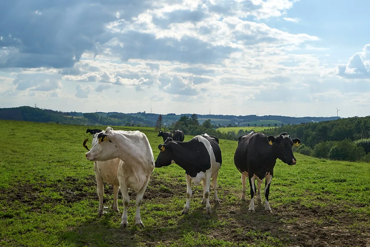
Thelaziosis in cattle: symptoms, treatment, and prevention
Thelaziosis is a dangerous ocular helminthiasis in cattle, transmitted by pasture flies during the summer period. The disease is caused by small nematodes that localize in the conjunctival sac, under the third eyelid, in the ducts of the lacrimal gland, and the nasolacrimal duct. Due to itching and pain, the animals become restless, shake their heads, and lose their appetite, which leads to a sharp drop in milk yield.
The disease can be recognized by characteristic clinical signs. Affected animals exhibit lacrimation, photophobia, redness and edema of the conjunctiva, and swelling of the eyelids. In neglected cases, keratitis, corneal ulcers, and corneal opacity develop. The disease proceeds acutely, especially in calves aged 4 months and older.
To make an accurate diagnosis, veterinary specialists rely on clinical signs and examine swabs from the conjunctival cavities of the animals. Timely detection of the pathogen prevents severe damage to the cornea. Once the disease is confirmed, comprehensive therapy is initiated immediately.
- Length of pathogen nematodes — up to 21 mm
- Development of larvae in flies — 3–4 weeks
- Maturation period in cattle eye — 125–200 days
- Duration of the disease — 1–2 months
| Washing solution | Concentration / Formulation |
|---|---|
| Boric acid solution | 2–3 % |
| Lysol emulsion | 3 % |
| Ichthyol emulsion | 3 % |
| Aqueous iodine solution (heated to 38–39 °С) | 1 g of crystalline iodine, 2 g of potassium iodide, 2 l of boiled water |
- Wash the animal's ocular cavity with one of the therapeutic solutions using a syringe with a soft tip under moderate pressure.
- Repeat the washing three times at intervals of 2–3 days.
- In case of complication with a secondary infection (purulent conjunctivitis, keratitis), prescribe symptomatic treatment with penicillin or sulfonamide preparations.
- Perform systemic deworming: administer doramectin (subcutaneously or intramuscularly at a dose of 0.2 mg/kg) or diethylcarbamazine (subcutaneously at a dose of 9–13 mg/kg).
Preventive deworming of the entire herd is carried out during the housing period or in the spring before the flight of face flies begins. After the first treatment, the procedure must be repeated every 7–8 days.
Trichinellosis: biology of the parasite and economic risks
Trichinellosis is an acutely or chronically occurring parasitic disease common to both animals and humans. The pathogen is a small nematode: the male has a length of 1.4–1.6 mm and a width of 0.14 mm. Among the pathogens, *Trichinella spiralis* (swine), trichinella from predatory animals of Eurasia, trichinella from African predators, as well as trichinella from the raccoon dog from Dagestan are described.
Adult parasites localize in the small intestine, and larvae localize in the striated muscles of the hosts. Pigs, dogs, wolves, foxes, cats, bears, rats, and mice are susceptible to the disease. Trichinellosis also affects wild boars, badgers, arctic foxes, ermines, minks, sables, polecats, hedgehogs, tigers, wild carnivores, and rodents. Helminthiasis is also recorded in marine mammals — whales, walruses, and seals.
Trichinellosis is extremely dangerous for humans; it proceeds severely and often results in death. Carcasses of infected animals are completely destroyed regardless of the intensity of the infestation, which causes huge economic damage to farms.
- Hosts of the parasite — more than 100 species of mammals
- Lifespan of females in the intestine — up to 8 weeks
- Infectivity of larvae — after 17.5 days
- Viability in muscles — up to 25 years
Infection of animals and humans occurs when eating meat containing live encapsulated trichinella larvae. In the body of a new host, the parasites undergo a complex cycle of development, sequentially changing location. The full path of the larva's transformation into an adult organism and the process of subsequent muscle infection consists of several key stages.
- In the host's stomach, the capsule of the ingested meat is destroyed, releasing a larva about 1 mm long, which quickly migrates to the small intestine.
- The parasites reach sexual maturity; already after 44 hours, mature eggs appear in the uterus of the females. Females embed their head end into the mucous membrane and are fertilized, after which the males die.
- On the 4th day after infection, the females begin to give birth to live larvae (one individual produces up to 2100 larvae, 1.2 mm in length and 0.006 mm in width).
- The larvae are carried by the blood and lymph flow throughout the body, penetrate under the sarcolemma of muscle fibers, and coil into a spiral.
- From the 4th to the 12th week, a capsule forms around the larvae. In pigs, it begins to calcify after 6 months, and this process is completely finished after 15–16 months.
Clinical picture, diagnostics, and treatment of trichinellosis in pigs
When affected by trichinellosis, the parasite larvae migrate throughout the pig's body, injuring tissues and causing internal hemorrhages. Their metabolic and decay products cause general intoxication, and adult individuals disrupt the integrity of the intestinal mucosa, opening the way for secondary infections. This leads to severe sensitization of the animal's body. The disease proceeds without specific clinical signs, but the veterinarian should pay attention to short-term temperature spikes, general depression, muscle pain, skin itching, and intermittent lameness.
Trichinella larvae are extremely resistant to external influences, so standard methods of meat culinary processing do not guarantee safety. They survive in lightly salted or standard smoked meat, and remain active for months in rotting meat. The parasite can only be destroyed in pork carcasses through strictly controlled temperature exposure.
| Environmental conditions and temperature regime | Survival time of Trichinella larvae |
|---|---|
| Rotting meat | Up to 4 months |
| Freezing to -10 °С | For a long time |
| Freezing to -30 °С | Die within 52–64 hours |
| Heating to 50 °С | Die within 15–18 hours |
A post-mortem diagnosis is established based on the results of carcass inspection. Most often, Trichinella localize in the pillars of the diaphragm, the muscles of the tongue, esophagus, larynx, as well as in intercostal and thoracic muscles. In rare cases, the parasites can be detected in parenchymal organs and the muscular layers of pork fat. Post-mortem diagnostics include mandatory trichinelloscopy: compression testing or the digestion method of samples in artificial gastric juice, which is considered the most accurate.
- Number of sections for analysis — 24 pcs.
- Size of section from the diaphragm pillar — size of a wheat grain
- Fenbendazole course for muscular form — 1–3 days
- Fenbendazole dosage — 2.5–50 mg/kg
No specific treatment for trichinellosis in animals has been developed, so the primary efforts of the veterinary service must be directed towards prevention. It is strictly forbidden to feed pigs slaughterhouse waste without prior decontamination. On farms, it is necessary to carry out rigorous rodent control, as mice and rats are the key reservoir of infection and actively carry it into holdings. For humans infected with trichinellosis, thiabendazole and other benzimidazoles are used.
Regardless of the intensity of Trichinella infestation, carcasses of pigs, wild boars, bears, and coypus are strictly prohibited for human consumption. All such carcasses are subject to technical disposal only, and it is forbidden on fur farms to feed untreated carcasses of game animals, dogs, and cats to livestock animals.
Fundamentals of Protozoology and Specifics of Piroplasmidoses
Protozooses are dangerous diseases of livestock animals and poultry caused by single-celled organisms (protozoa). Their study is covered by protozoology. Anatomically, protozoa consist of a nucleus and cytoplasm. They use flagella, cilia, or pseudopodia as organs of locomotion.
Nutrients are acquired either through a special organelle—the cytostome—or by absorption across the entire body surface via phagocytosis or pinocytosis. The reproduction of these organisms occurs through both asexual and sexual pathways. Asexual reproduction includes simple or multiple fission (schizogony), as well as budding. In the parasite life cycle, these processes regularly alternate with the sexual process—copulation or conjugation.
For veterinary medicine, the three classes of protozoa of greatest significance are Sporozoea, Flagellata, and Ciliata. The Sporozoea class unites several key orders that cause dangerous diseases in livestock animals and poultry. These include the orders Piroplasmida, Coccidiida, and Microsporidia.
- Order Piroplasmida (causative agents of piroplasmidoses);
- Order Coccidiida (causative agents of coccidioses);
- Order Microsporidia (causative agents of microsporidioses).
Piroplasmidoses, also known as hemosporidioses, are caused by sporozoans of the Babesiidae and Theileriidae families. These diseases are widely spread in southern regions. They cause serious economic damage to animal husbandry, which is driven by the reduction in the productivity of livestock animals and their mass mortality.
After recovery, animals develop non-sterile immunity lasting from 4 months to 2 years or more. During this period, animals remain carriers of the pathogen.
| Livestock animal mortality rate | Value |
|---|---|
| Mass mortality in piroplasmidoses | 20–60% or more |
| Mortality of adult animals in babesioses | 40–60% |
Piroplasmidoses pose a serious threat to livestock farms, causing mass death of livestock. The disease spreads rapidly, but only under the presence of three mandatory conditions: infected livestock animals (or latent carriers among the recovered stock), active ixodid tick vectors, and susceptible livestock. Without breaking this chain, it is impossible to stop the contamination of pastures.
Babesiosis: Disease Progression, Symptoms, and Treatment Regimen
Babesioses are acute protozoan diseases that affect cattle">cattle, sheep, and goats. Adult livestock animals suffer the most severe course of the disease, while young stock recover asymptomatically, remaining latent sources of infection. The disease pathogens (1.5–2.4 µm in size) penetrate red blood cells, destroy them, disrupt oxidative processes in the body, and cause general intoxication. Tick vectors become infected while ingesting blood, multiply the parasites in their bodies via multiple fission, and transmit them to the next generation transovarially (through eggs). Livestock animals are primarily infected on forest and shrub-covered pastures, but ticks can also be brought into stalls along with freshly cut grass.
- Mortality of adult animals — 40–60%
- Incubation period of the disease — 8–14 days
- Critical body temperature — 40–42 °С
- Decrease in red blood cells — down to 2 million per 1 mm³
- Drop in hemoglobin — down to 30%
The first signs of infection manifest as lethargy, loss of appetite, cessation of rumination, and gastrointestinal atony, which progresses to constipation. In dairy cows, milk yield drops sharply, while pulse and respiration rates increase. By the 2nd–3rd day, urine becomes pink, then dark red (in goats, urine color does not change). Mucous membranes first turn pale, and then become jaundiced.
In the absence of treatment or if it is started untimely, sick livestock animals die on the 4th–8th day of the disease.
Upon detection of symptoms, it is necessary to immediately isolate the sick and begin specific drug therapy. Recovered livestock acquire non-sterile immunity, which persists for up to one year. Timely intervention allows for saving the majority of the herd and preventing severe complications.
| Specific drug | Concentration and dosage | Administration method |
|---|---|---|
| Hemosporidin | 1% solution, 0.5 mg/kg | Subcutaneous or intramuscular |
| Flavacridine | 1% solution, 3–4 mg/kg | Intravenous |
| Piroplasmin | 1% solution, 2 mg | Subcutaneous |
| Berenil (azidine) | 7% solution, 3.5 mg/kg | Intramuscular |
If necessary, the therapeutic treatment is repeated after 24–48 hours. Additionally, caffeine is prescribed to the animals, laxatives are given, and the diet must include succulent feed, green grass, and milk or skimmed milk at 1.5–2 liters 2–3 times a day.
Prevention of babesiosis consists of pasture cultivation and tick eradication. In case of an outbreak on a farm, chemoprophylaxis is used. For this purpose, azidine is administered to the entire livestock population at a therapeutic dosage once every 12 days.
Piroplasmosis: diagnosis and progression characteristics
Piroplasmosis has an acute course and affects not only cattle">cattle, but also sheep, goats, horses, mules, dogs, and pigs. The causative agents are larger than Babesia (2–6 µm) and are localized in the center of red blood cells, affecting up to 20% of all blood cells. In the bodies of animals, the parasites multiply by division or budding, and in tick vectors, they undergo a cycle of schizogony. The spread of the disease depends on the climate: in the south of Kazakhstan, ticks are active almost year-round, while in northern regions, outbreaks are strictly seasonal (spring, summer, and autumn). Infection occurs in pastures, as well as in stalls when ticks are brought in with grass.
- Assess the epizootological situation: presence of active tick vectors on the animals' bodies, season of the year, and type of pastures.
- Conduct a clinical examination: record fever (41–42 °С), depressed state, dyspnea, jaundice of the conjunctiva, and atony of the forestomachs.
- Check for characteristic symptoms: appearance of hemoglobinuria (red urine) on the 2nd–3rd day in ruminants or cloudy urine in horses.
- Take blood smears for microscopic examination to detect piroplasms in red blood cells.
- In case of death, perform a necropsy to confirm the diagnosis by spleen enlargement and jaundice of internal tissues.
- Mortality of adult animals — up to 60%
- Incubation period in cattle — 14–24 days
- Incubation period in sheep and horses — 8–10 days
- Duration of the disease in horses — 8–12 days
The clinical course of piroplasmosis has its own peculiarities in different animal species. In sheep and cows, death occurs rapidly without treatment — on the 5th–7th day of the disease. In horses, the disease lasts slightly longer (8–12 days), is accompanied by the discharge of cloudy jaundiced urine, and pregnant mares abort when infected. Timely diagnosis and isolation of affected individuals help to avoid mass mortality on the farm.
Specific therapy for piroplasmosis and prevention of tick-borne infestations
Upon confirmation of piroplasmosis in cattle, treatment must begin immediately, as delay leads to severe productivity losses and animal death. Therapy must be comprehensive: in addition to specific antiparasitic drugs, supportive cardiac and laxative agents must be prescribed. Sick livestock are moved to a gentle diet, including fresh green grass, chopped root crops, as well as warm whole milk or fresh skimmed milk.
- Dosage of trypansin — 5 mg/kg in a 1% solution intravenously
- Dosage of berenil (azidine) — 3.5 mg/kg in a 7% solution intramuscularly
- Dosage of trypaflavine — 3–4 mg/kg in a 1% solution intravenously
When treating with berenil (azidine) or trypaflavine, the drugs are administered once or twice with a mandatory 24-hour interval. Other animal species require their own treatment regimens: azidine or hemosporidin is prescribed for sheep, and flavacridine or azidine for horses.
Prevention of piroplasmosis is based on the destruction of tick vectors in the environment and directly on the animals. Agrotechnical measures are carried out in fields — ploughing pastures followed by the sowing of cultivated grasses. Livestock and livestock facilities are regularly treated with acaricides, and if necessary, chemoprophylaxis with azidine is used.
Eimeriosis in livestock animals: pathogen life cycle, symptoms, and treatment
Coccidiosis poses a serious threat to livestock farms, especially for young animals. These diseases are caused by protozoa of the Eimeriidae family; depending on the genus, these include eimeriosis, isosporosis, toxoplasmosis, and other pathologies. Eimeriosis, which affects the intestines, liver, and other internal organs of cattle, sheep, rabbits, and poultry, is of the greatest practical importance.
The disease is caused by microscopic Eimeria ranging from 11 to 40 μm in size. Parasites are localized primarily in the intestinal epithelium, although in rabbits they also affect the bile ducts of the liver, and in poultry — the mucous membrane of the renal pelvis. The parasite's life cycle includes asexual reproduction (schizogony) and sexual reproduction (gametogony) within the host organism, as well as sporogony in the external environment.
Under favorable conditions (warmth, humidity, access to oxygen), sporogony in the external environment is completed within 2–3 days. At this stage, the oocyst cytoplasm divides into 4 sporoblasts, rendering it infective. Such sporulated oocysts can remain viable in the environment for a year or more.
| Stage on the diagram | Process of pathogen (Eimeria) development |
|---|---|
| 1 | Sporozoite |
| 2–4 | Development of first and subsequent generation schizonts |
| 5 | Third-generation merozoites |
| 6 | Development of microgametes |
| 6а | Development of macrogametes |
| 7 | Microgametes |
| 8 | Zygote (oocyst) |
| 9 | Non-sporulated oocyst released from the rabbit's body |
| 10–12 | Sporogony in the external environment |
| I–III | Asexual reproduction (schizogony) |
| IV | Gametogony |
The main sources of infection are sick animals and asymptomatic carriers. The spread of the infestation is facilitated by a warm, humid climate, overcrowding, poor-quality feeding, mixed-age group housing, and infrequent removal of manure. By penetrating the epithelial cells of internal organs, the parasites destroy them, which leads to digestive disorders, urinary system failure, and severe intoxication.
The incubation period in cattle and sheep lasts 2–3 weeks, and in rabbits and poultry — from 4 to 12 days. In acute cases, young animals exhibit depression, loss of appetite, fever with a temperature rise of 1–2 °C, as well as diarrhea containing mucus and blood. By the 2nd–3rd day, feces darken, anemia and jaundice develop; in rabbits and chicks, limb paralysis is possible, leading to death within 2–10 days. In the chronic form, symptoms are milder: animals lag in growth, diarrhea alternates with constipation, but animals that have recovered remain carriers of the infection for a long time.
The diagnosis is made comprehensively based on symptoms, epizootiological data, and microscopy of feces or scrapings from affected organs. Sick individuals are immediately isolated for treatment. For therapy, animals are prescribed sulfadimezin, norsulfazol, sulfadimethoxine, iramine, coccidin, or other similar agents.
Eimeria oocysts are extremely resistant in the external environment. For the disinfection of premises, a 7% ammonia solution or a hot 10% iodine monochloride solution must be used, and all manure must undergo biothermal decontamination.
To prevent coccidiosis, young animals must be kept in clean, dry housing separated from adult livestock. For this purpose, rabbits and chicks are placed in cages with wire-mesh floors. In farms affected by coccidiosis, timely chemoprophylaxis is mandatory.
Drugs (coccidin, sulfonamides, and other coccidiostats) are administered in a mixture with feed or water. They must be alternated regularly, otherwise, the pathogens quickly develop resistance to the active ingredients.
Psoroptic mange in animals: pathogen biology, symptoms, and treatment regimen
Psoroptic mange (common mange) is a widespread disease of sheep, cattle, and horses, caused by acariform mites of the genus Psoroptes. The disease is accompanied by intense skin itching, dermatitis, hair loss, and general exhaustion of the body. Humans do not contract this form of mange. Infection occurs through direct contact between healthy and sick animals, as well as through grooming tools and the clothing of staff. The spread of infestation is facilitated by overcrowding, high humidity in premises, inadequate feeding, and secondary diseases.
The pathogens are localized on the surface of the skin. These are dioecious mites without eyes, with a body length of up to 0.8 mm and a proboscis adapted for piercing the epidermis and sucking lymph. On the ventral side of the pest, there are 4 pairs of legs, of which 3 pairs are equipped with suckers: females have suckers on the 1st, 2nd, and 4th pairs of legs, while in males, the suckers on the 4th pair are undeveloped. Females lay eggs directly on the animal's skin.
- Egg hatching time — 3–7 days
- Development of the new generation — 2–3 weeks
- Life span of the mite — up to 60 days
- Number of eggs per female — about 60
- Viability in premises — up to 2 months
- Death at temperatures below 5 °C — within 5–9 days
Favorable conditions for the development of parasites are air humidity of 85–90% and a temperature of 36–37 °С. The larvae molt twice, passing through the protonymph and teleonymph stages, after which they turn into adults. The disease progresses most intensively during the autumn-winter period.
The incubation period of the disease lasts from 14 to 60 days, transitioning into an acute or chronic form. The main symptom of psoroptosis is severe itching, which becomes especially intense at night, after driving or after being caught in the rain. The wool in the affected areas becomes ruffled, is easily pulled out or falls out spontaneously, after which crusts form on the skin and it thickens. With extensive damage, the livestock begins to lose weight, and in the summer, the signs of the disease weaken and it enters a latent course.
The localization of the lesions depends on the species of the livestock animal. In sheep, the back, sacrum, and sides are affected. In cattle, mites nest at the base of the horns and on the upper part of the neck. In horses, the affected skin areas include the neck, under the mane on the back, the poll, and the base of the tail.
The diagnosis is established based on clinical symptoms and mandatory microscopy of skin scrapings. For analysis, material must be taken strictly at the border between the affected and healthy skin areas. Upon confirmation of the disease, quarantine restrictions are imposed on the farm.
Specific control measures depend on the season of the year. In the warm season, animals are bathed or sprayed with a neostomazan emulsion. If mange is detected in the winter period, the flock is injected with Ivomec or another ivermectin-based preparation.
Read next
Veterinary medicine For students
Veterinary and sanitary examination of meat in cases of cysticercosis and disease prevention
Veterinary medicine For students
Characteristics of sarcoptic mange and demodicosis in livestock animals
Veterinary medicine For students