
Veterinary and sanitary examination of meat in cases of cysticercosis and disease prevention
18 min read
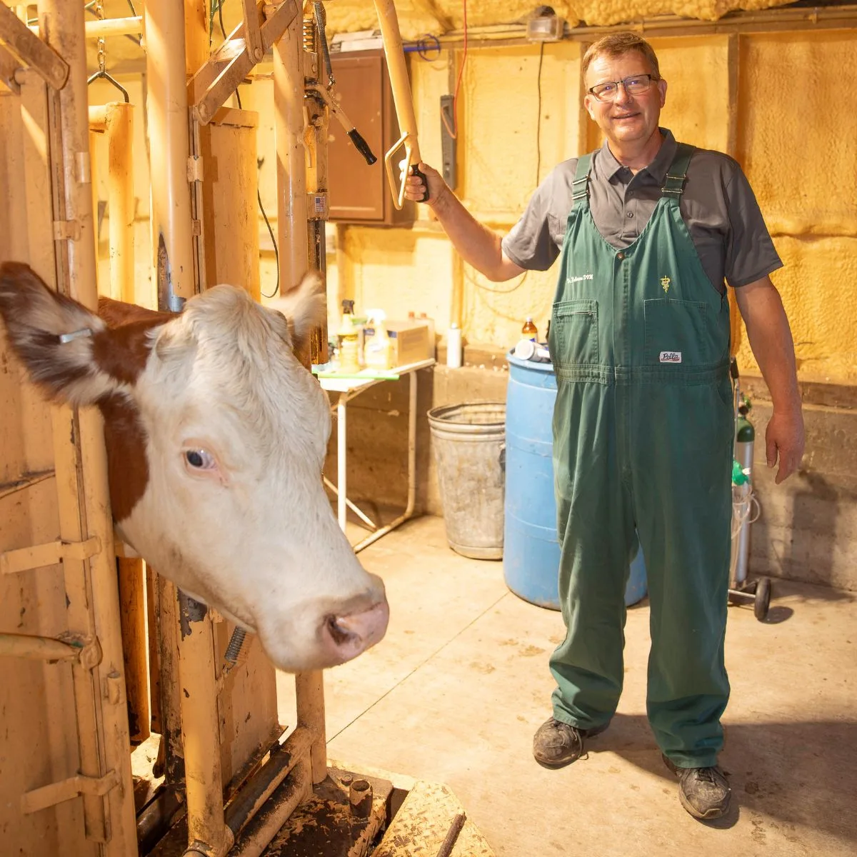
Finnosis (cysticercosis): veterinary and sanitary examination rules for carcasses
The control of cysticercosis requires close cooperation between veterinary and medical services, since humans are the only definitive host of the parasite and the source of infection for livestock animals. Thorough examination of cattle and pig meat at slaughterhouses allows for the timely detection of infestation. This prevents the infection of humans with dangerous diseases — taeniasis and taeniarhynchosis, and also prevents the possibility of infecting the animals themselves.
To detect cysticerci, a veterinary specialist performs a detailed inspection of the carcass. The examination of muscle tissue is carried out in a strictly defined sequence. Particular attention is paid to the areas where parasites are most frequently localized.
- Incision the external and internal masseter muscles, and also examine the heart tissues.
- If cysticerci are detected, make two additional parallel incisions: in cattle — in the neck muscles, deep lumbar muscles, and diaphragm; in pigs — in the lumbar muscles, occipital muscles, and diaphragm.
- Assess the density of parasite tissue infestation over an area of 40 cm² and determine the further procedure for handling the meat.
| Number of cysticerci | Detection conditions | Examination result and follow-up procedure |
|---|---|---|
| More than three | In an area of 40 cm² of head muscles, heart, and in one of the carcass muscle incisions. | The entire carcass (except the intestines) is sent for technical disposal or destroyed. Internal fat is rendered for food purposes. |
| More than three living or dead | In an area of 40 cm² of head and heart muscle incisions, while there are no cysticerci in other incisions of the aforementioned muscles or no more than three are found. | The head and heart are sent for disposal. The carcass and other organs (except the intestines) are decontaminated by boiling, freezing, or salting, followed by use for the preparation of minced sausage products or minced meat canned goods. Decontaminated by-products are sent for industrial processing; internal fat is rendered. |
All cases of cysticercosis detection in animals must be immediately reported to the appropriate medical organizations. This is necessary for examination and the implementation of therapeutic and preventive measures among persons who were the source of infection for the animals.
Basic preventive measures include systematic examination of humans to identify all those infected with taeniasis and taeniarhynchosis, followed by their mandatory deworming. It is necessary to protect soil and water bodies from human fecal contamination, install public and individual toilets, and conduct sanitary control over excrement disposal and the state of sewage. It is important for people to follow personal hygiene measures and not to consume raw, undercooked, or underfried meat and internal organs.
Echinococcosis: pathogen biology and farm control
Echinococcosis is a dangerous anthropozoonosis, which usually proceeds asymptomatically in sheep, goats, cattle, pigs, camels, deer, and less often horses and other mammals. Humans also suffer from this disease, sometimes with fatal outcomes. The chronic course of the infestation significantly reduces animal productivity, delays the growth and normal physiological development of young stock, and reduces the body's resistance to other diseases.
The disease is caused by the larval stage of a cestode localized in the liver, lungs, and less often in other organs and tissues. The pathogen is a single-chamber cyst filled with liquid, the size of which varies from barely visible to the size of a newborn baby's head. The tapeworm stage of the echinococcus parasitizes the small intestine of dogs, wolves, jackals, and less often foxes, while a mature proglottid usually exceeds the rest of the strobila in length.
- Length of the tapeworm stage strobila — up to 6 mm
- Number of proglottids in the strobila — 34 units
- Number of eggs in a mature proglottid — about 800 units
- Development of the cyst to the invasive stage — not earlier than 6 months
- Parasite development period in dogs — 2–3 months
- Lifespan of echinococcus in dogs — 5–6 months
Definitive hosts shed mature echinococcus proglottids into the external environment along with excrement; these proglottids are actively moving, spreading up to 15–20 cm and climbing up plant stems. During movement, their walls rupture and eggs are released. Intermediate hosts become infected by ingesting oncospheres or parasite proglottids with feed or water. In the stomach, the oncosphere shells are destroyed, the embryos penetrate the intestinal wall and are carried by blood or lymph into organs, where they slowly form into a cyst that lives for many years.
During veterinary inspection, organs affected by echinococcosis, and sometimes entire carcasses, are destroyed. Do not allow dogs to eat affected organs (most often the lungs and liver) of fallen or slaughtered livestock — this is the main route of infection for carnivores.
Sheep are of the greatest importance in the epizootiology and epidemiology of echinococcosis, and dogs (especially stray and shepherd dogs) and wild predators serve as the source of infection. The spread of the infestation is facilitated by poor feeding of livestock animals and unsatisfactory veterinary and sanitary conditions on the farm. Humans become infected through direct contact with infested dogs, as well as by eating vegetables and fruits contaminated with echinococcus eggs.
Figure 34 - Biology A – intermediate hosts; B –
Pathogenesis. Larvae of echinococcus exert mechanical, toxic, and allergic effects. Depending on the localization, number, and size of the echinococcal cysts, various local and systemic disorders arise in the body of the intermediate host.
Symptoms of the disease. For a long time after infection, echinococcosis proceeds without pronounced signs. Later, clinical signs gradually appear, which are quite diverse in echinococcosis.
In most cases, severe emaciation occurs, leading to a decrease in the productivity of animals; in sheep, the fleece becomes ruffled and often falls out, cows show a decrease in milk yield. When the liver is affected, digestive disturbances develop, the area of hepatic dullness increases, and palpation of this area is painful. In pulmonary echinococcosis, symptoms include coughing and labored breathing.
Pathoanatomical changes. Echinococcal cysts are usually found in the liver and lungs, less often in the kidneys and spleen; sometimes they are located near the surface of the organ, protruding above its serous membrane.
Diagnosis. It is difficult to diagnose echinococcosis based on the clinical picture; in Kazakhstan, allergic and immunological methods are being developed.
Treatment of animals suffering from larval echinococcosis has not been developed. However, relatively recently, two groups of substances were synthesized: isoquinoline pyrazine derivatives and benzimidazole derivatives.
The first group includes praziquantel (Droncit). This drug is successfully used for the treatment of cysticercosis in ruminants. The drug is administered orally, subcutaneously, and intramuscularly once at a dose of 5 mg/kg.
The second group of anthelmintics, tested with positive results for larval cestodiasis, includes the following dosages:
| Drug | Dose (mg/kg) |
| Mebendazole (orally for dogs) | 60-100 |
| Fenbendazole | 50 |
| Cambendazole | 20 |
Prophylaxis is, in principle, the same as for other larval taeniid infections.
Nematodiasis — diseases caused by helminths from the class of round parasitic worms — nematodes. This is the largest group of helminthiasis in the entire field of veterinary helminthology.
Anatomical and morphological characteristics of nematodes. Nematodes are characterized by an elongated, thread-like or spindle-shaped body. The body length of various nematode species ranges from 1 mm to 10 m. Externally, the body is covered with a dense layer of cuticle, which, together with the underlying muscle tissue, forms the so-called skin-muscle sac, in which the internal organs are located. The surface of the cuticle is uneven; it can be striated in various directions, and some nematode species have cuticular structures of various shapes - plates, ridges, and spines, which serve to anchor the nematodes. The digestive system consists of an esophagus, originating from the mouth opening at the anterior end, and an intestine located along the entire length of the body; near the posterior end of the body is the anal opening. The excretory system is represented by tubules; they begin in the posterior part of the body and then merge into a single canal that opens in the anterior part of the body. The nervous system consists of a central nerve ring surrounding the esophagus, with nerve trunks extending from it, and a number of nerve ganglia. With rare exceptions, all nematodes are dioecious animals; as a rule, females are significantly larger than males. The female reproductive apparatus is represented by two ovaries, two uteri (rarely one or many) with oviducts, and one vagina, which opens on the ventral side of the body in the form of a genital slit (vulva), closer to the head or tail end. The male reproductive apparatus consists of a testis and a highly convoluted vas deferens, consisting of a seminal vesicle and an ejaculatory duct. The vas deferens opens into the anal opening of the intestine — the cloaca. Near it, in some nematode species, there is a complex of auxiliary male reproductive organs. Of these, spicules are of great importance — during copulation, the spicules are inserted into the vulva, fixing the female and widening the vagina; semen flows along them into the female reproductive tract.
The biological cycle of nematode development is characterized by great diversity. As among all other parasitic worms, nematodes are divided into geohelminths, which develop directly without the involvement of intermediate hosts, and biohelminths, whose developmental cycle occurs with the mandatory participation of intermediate hosts. In addition, many nematode species have reservoir hosts. Females of certain nematode species release eggs or larvae through the genital opening, and accordingly, they are called oviparous and viviparous. Eggs contain either a fully formed larva or individual blastomeres.
How ascariasis develops and why larval migration is dangerous
Nematode larvae reach the infective stage after two molts in the external environment for geohelminths or inside an intermediate host for biohelminths. In total, representatives of eight suborders of parasitic nematodes are of veterinary importance. In the direct path of development, livestock animals become infected by ingesting eggs or larvae with feed and water, while pathogens such as Strongyloides, hookworms, and Uncinaria can actively penetrate even through intact skin. Inside the host, larvae develop either directly in the digestive tract (such as Trichocephalata and Oxyurata) or after a complex migration through tissues and organs (such as Strongylata and Ascaridata).
- Age of highest susceptibility — up to 6–7 months
- Daily productivity of one female — up to 200 thousand eggs
- Optimal temperature for egg development — 20–30 °С
- Larval maturation period in the environment — 2–3 weeks
- Development to the sexually mature stage — 1.5–2.5 months
The causative agent of porcine ascariasis is a large white nematode, the head end of which is equipped with three lips. The males of this parasite reach 10.5–22 cm in length, and females grow up to 23–30 cm. Infection of the herd occurs mainly directly in the pigsty. The parasite larvae affect lymph nodes, lungs, and liver, causing serious allergic changes: eosinophilic infiltrates, bronchopneumonia, and pleurisy.
- The piglet ingests infective eggs, and larvae hatch in its intestine.
- The parasites penetrate the intestinal wall and migrate through the portal vein to the liver, destroying its cells.
- Through the inferior vena cava, the right heart, and the pulmonary artery, the larvae penetrate the lung capillaries.
- Tearing the blood vessels, the parasites exit into the pulmonary alveoli, bronchioles, bronchi, and trachea.
- The larvae are coughed up into the oral cavity, re-ingested, and develop into adults in the small intestine.
In the intestine, ascarids are held in place by an arc-shaped curvature of the body, bracing their ends against the walls and resisting peristalsis. They feed on intestinal contents and live there for 7 to 10 months, after which they are spontaneously expelled. The number of simultaneously parasitizing individuals in the intestine can vary from individual specimens to several hundreds or thousands.
Clinical signs, diagnosis, and treatment regimens
Symptoms of the disease directly depend on the developmental phase of the helminths. The disease is most severe in suckling piglets and young stock, as larval migration causes them acute allergic and mechanical tissue damage. Adult pigs rarely get sick and carry the infestation asymptomatically, while remaining constant sources of infection for the farm.
In the first days after infection, larval migration provokes acute ascaridic pneumonia. In piglets, body temperature rises to 41.5 °С, respiration quickens, and coughing, dyspnea, salivation, and vomiting appear. The disease lasts from 6 to 15 days and can be accompanied by an allergic skin rash, hives, papules, and periodic convulsions.
An intravital diagnosis of ascariasis is based on laboratory examination of pig feces. Posthumously, infection is determined by finding adult ascarids in the intestine. Additionally, pieces of lung and liver tissue are examined to detect migrating larvae.
With age, the susceptibility of pigs to ascariasis decreases significantly due to the formation of acquired immunity.
For deworming of pigs, individual or group administration of anti-parasitic drugs with feed or water is used. Dosages of active ingredients are calculated in accordance with the recommendations of a veterinarian.
| Drug | Dosage |
|---|---|
| Mebendazole | 20 mg/kg |
| Oxybendazole | 10–15 mg/kg |
| Fenbendazole | 10–15 mg/kg |
| Levamisole (internal) | 7.5 mg/kg |
| Piperazine | 300 mg/kg (up to 15 g) |
| Ivermectin (internal) | 0.3 mg/kg |
| Hygromycin B (premix) | 8–12 g per ton of feed |
| Sodium silicofluoride (3 days) | 1.5–2.5 kg per ton of feed |
Prevention. In farms affected by porcine ascariasis, scheduled preventive deworming is carried out annually. Sows are dewormed a month before mass farrowing, before being moved to open-air enclosures, and in autumn before being moved to stall housing.
Young stock are assigned deworming according to the following schedules:
- From April to December: 35-40 days, 50-55 days, and 90 days.
- From December to April: 50-55 days and 90 days.
After deworming, before each farrowing cycle, or in fattening pigsties — during the period of pig re-sorting, before loading the pens, disinfection of premises is carried out with hot ash lye, hot solutions of caustic soda or potash, a 10-20% suspension of freshly slaked lime, or a 10% hot emulsion of xylonaphth.
Manure is removed daily and transported to a manure storage area for biothermal disinfection. Housing and proper feeding of gestating sows and piglets are of important preventive significance.
Parascaridosis — a disease of equines: horses, donkeys, mules, caused by nematodes parasitizing the small intestine, the larval forms of which migrate in the body via the hepato-pulmonary pathway.
Pathogen. A white nematode with an elastic, spindle-shaped body. The male is 15—28 cm long, differing in appearance from the female by its smaller size and curved tail end. The female reaches 37 cm in length.
Biology of the pathogen. The life cycle is direct, following the ascarid type. Larvae emerge from the egg in the horse's intestine, where they are ingested along with contaminated feed and water. Through the intestinal mucosa, they penetrate the blood vessels, then travel through the liver and the right side of the heart to the lungs, developing there for some time before migrating into the alveoli, bronchi, and trachea, from where they are coughed up into the oral cavity. From the latter, the larvae are swallowed again and end up a second time in the horse's intestine, where they grow into sexually mature helminths. The development time for parascarids from the moment of egg ingestion to sexual maturity is 2—2.5 months. Parascarids parasitize in the intestine for up to a year or more.
Figure 35 – Biology Examine the developmental stages of the pathogen.
Epizootiological data. Parascaridosis is an invasive disease that primarily affects young animals under one year of age. The infestation is particularly severe, with fatal cases, in suckling foals, which can become infected in the first days and weeks of their lives. Adult horses are also heavily infected (up to 46-50%), but they are generally asymptomatic carriers of parascarids. Poor-quality feeding reduces the body's resistance and therefore contributes to more intensive infection of foals.
The source of infestation is infected livestock animals, which shed parascarid eggs into the external environment along with feces, contaminating the surrounding territory and grooming equipment:
- stalls;
- feeders;
- shovels;
- brooms, etc.
The most favorable conditions for parascaridosis infection in the stable and on the pasture are from May to September. Therefore, the extensiveness of equine parascaridosis begins to increase from May — June, reaching a maximum in September — October and remaining at a high level until January — February.
The symptoms of the disease are quite varied and depend significantly on the age of the animals and the intensity of the infestation. Adult animals are generally parasite carriers, so the disease in them proceeds asymptomatically.
In foals, at the beginning of the disease — during the larval migration period — enteritis and diarrhea (3—4 days) are noted. After this, signs of bronchopneumonia develop: a short-term rise in temperature, coughing, rapid breathing, and serous-mucous nasal discharge. Sometimes there are episodes of nervous agitation. These phenomena are most pronounced on the 9—16th day after infection and usually last 4—7 days, then gradually disappear.
The disease in the stage of mature parascarid parasitism is characterized by gradual emaciation of the affected animals, periodic diarrhea alternating with constipation; foals lag in growth, their abdomen size increases, and sometimes they experience colic.
Diagnosis during the animal's life is based on coprological studies using the Fülleborn method. Sometimes parascarids are shed with feces; the passage of these helminths can be induced by diagnostic deworming. Post-mortem, the disease is confirmed by detecting parascarids in the intestine.
Treatment. The following drugs are used:
| Mebendazole | oral 6-8 mg/kg |
| Thiabendazole | oral 100 mg/kg |
| Fenbendazole (Panacur) | oral 7.5-10 mg/kg |
| Febantel | 650 mg/kg |
| Piperazine | oral, after a fasting diet at a dose of 40-50 mg/kg |
| Ivermectin | oral 0.2 mg/kg |
| Morantel | 10 mg/kg |
Prevention. In farms affected by parascaridosis, mandatory deworming of horses is carried out at the following times: for foals of the current year of birth — the first time in August, the second time — after weaning; for young animals 1—2 years old and adult horses — in March — April and in October — November.
In horse-breeding farms with herd management, suckling foals starting from 3 months of age are fed 10 g of piperazine per dose via a group method for 2 consecutive days each month. From August until the end of the year, the dose is increased to 15 g per dose; this drug is administered for 2 consecutive days every 2—3 months. Horses at racetracks are dewormed with piperazine every 2—3 months.
After medical treatments, it is necessary to disinfect the areas where the animals are kept immediately. If deworming took place directly on the pasture, it is important to strictly observe the rules for herd isolation. In affected farms, a complex of sanitary measures similar to the control measures for swine ascaridosis is additionally recommended.
- Perform animal treatment with the drugs.
- Transfer the horses to an isolated plot of pasture and keep them there for 3–4 days.
- After this period ends, completely plough the used isolated plot.
- In parallel, carry out thorough disinfestation of the stables.
Ascaridiosis of poultry: biology of the pathogen and control measures
Ascaridiosis is a widespread helminthiasis of poultry caused by nematodes that parasitize the small intestine. Chickens and young birds under 8–10 months of age are most susceptible to the disease, whereas adult individuals usually act as asymptomatic parasite carriers. The pathogen is dangerous not only for chickens: it affects turkeys, geese, guinea fowl, peacocks, pheasants, and capercaillies. Male parasites reach a length of 26–70 mm, females — 65–110 mm.
Infection occurs upon ingestion of invasive eggs with feed, water, or by consuming earthworms, which act as reservoir hosts. The peak of infestation occurs during the wet and warm season: infection levels rise in September — October, reach a maximum in November — January, and remain at a high level until May. In damp and shaded areas of outdoor runs, helminth eggs survive significantly longer than under direct sunlight, where they perish quickly. Transitioning poultry to cage housing almost completely eliminates the risk of infection.
- Larval development period in the external environment — 15–20 days
- Larval stay in the duodenal mucosa — about 9 days
- Release of young ascaridia into the intestinal lumen — on the 38th day
- Development period of the parasite to sexual maturity — 28–56 days
- Lifespan of the nematode in the intestine — 9–14 months
- Maximum fecundity of a single female — up to 50 million eggs
The natural resistance of poultry to ascaridiosis decreases sharply due to vitamin A deficiency in the diet, overcrowded housing, and unbalanced feeding.
Symptoms of the disease manifest on the 7th–10th day after infection. The sick bird becomes lethargic, inactive, sits ruffled with drooping wings, and its visible mucous membranes and comb turn pale. Alternating diarrhea and constipation, as well as mucus discharge from the beak, may also be observed. For ante-mortem diagnosis, coprological examinations using the Fülleborn or Darling methods are used, while post-mortem diagnosis is established based on the results of finding ascaridia in the intestine.
The main rule of prevention is isolated rearing of young stock. Keep chicks in brooders or acclimatizers and rotate outdoor pens every 1–2 years, preventing poultry from accessing areas where an infected flock was kept in the previous year.
| Drug | Dosage | Method and scheme of application |
|---|---|---|
| Mebendazole | 40 mg/kg | Oral |
| Fenbendazole | 10–40 mg/kg | Oral |
| Flubendazole | 5 mg/kg | Oral |
| Levamisole | 20 mg/kg | Oral |
| Piperazine | 100–250 mg/kg | Oral |
| Hygromycin B | 8–12 g of hygromycin per 1 ton of feed | As a premix with feed |
| Bithionol | 2–3 g/kg | Oral, twice at 1-day intervals |
Read next

Veterinary medicine For students
Thelaziasis in cattle: symptoms, diagnostics, and treatment
Veterinary medicine For students
Etiology of brucellosis and features of the infectious process in livestock animals
Veterinary medicine For students