
Etiology of brucellosis and features of the infectious process in livestock animals
18 min read
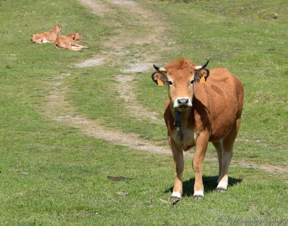
Brucellosis is a chronic infectious disease manifesting as abortions, retention of the placenta, endometritis, and reproductive disorders in livestock animals.
Pathogen. The genus includes 6 species: Br. including 9 biovars, 3, 4, and Br. canis — one each. All Brucellae are polymorphic; coccoid, ovoid, and rod-shaped forms are encountered.
The resistance of Brucellae to physical and chemical factors is low. At 60°C, they perish in 30 minutes, at 70°C — in 5-10 minutes, and at 90-100°C — instantly. In souring and chilled milk and cream, the microbe survives for up to 4-7 days; on clothing — up to 14 days; in cheeses, butter, brynza, and salted hides — up to 67 days; in salted meat — up to three months; in frozen meat and on wool — up to five months. In soil, water, manure, and roughage, the pathogen can remain viable for up to four months. Direct sunlight kills them in 3-4 hours, 1% solutions of creolin, phenol, or formaldehyde kill them in an hour, and 5% freshly slaked lime kills them in 2 hours.
Epizootiological data. Cattle , sheep, goats, pigs, deer, marals, yaks, buffaloes, horses, camels, dogs, cats, hares, saiga antelopes, foxes, rodents, and wild boars are susceptible to brucellosis .
In cattle, camels, and horses, brucellosis is caused by Br., in pigs by Br., in goats and sheep by Br., in dogs by Br., and possibly also by Br. and Br. Among cattle, sheep, goats, and pigs, brucellosis occurs in the form of epizootic outbreaks, while in horses, dogs, and other animals, it occurs as sporadic cases.
The source of the infectious agent is animals sick with brucellosis. A large number of pathogens are shed by animals through amniotic fluid, fetal membranes, and aborted fetuses. Infection occurs via the alimentary route (at any age) and through contact (in adult animals — via the genital route).
Course and symptoms. The incubation period lasts 15-30 days. If there are no pregnant animals among the susceptible livestock, the disease proceeds asymptomatically (latent form). In pregnant animals, brucellosis is characterized by abortions in the second half of pregnancy. Cows usually abort at 5-8 months, and sheep and goats — at 3-5 months of pregnancy. Sows can abort in both the first and second half of pregnancy, and dogs at the 40-50th day. In cattle and sheep, repeat abortions are rarely observed, while in pigs, they can be multiple. 1-2 days before the abortion, the udder swells, the external genitalia become inflamed, and a slight discharge of brownish-red mucous fluid from the vagina is noted. Abortions are accompanied by retention of the placenta and the development of endometritis. Brucellosis can be accompanied by serous bursitis, arthritis, and tendovaginitis, and in males — by orchitis and epididymitis.
Diagnostics. Regular (twice a year) serological tests are conducted. For mass prophylactic and diagnostic testing of livestock for brucellosis, the following methods are widely used:
- AG (agglutination test);
- CFT (complement fixation test);
- Rose Bengal test (RBT);
- Coombs test;
- Milk ring test (MRT);
- R-CFT (reaction of complement fixation with dilution).
Allergic tests have the greatest diagnostic value in the late stages of the disease. For these purposes, brucellin is used. The preparation is administered palpebrally under the skin of the lower eyelid in accordance with the following dosages:
| Animal species | Dose of brucellin |
| Sheep, goats, and deer | 0.5 ml |
| Cattle and buffaloes | 1 ml |
The reaction is assessed after 36-48 hours. Inflammation at the site of allergen administration is considered a positive reaction; animals are declared sick and subject to slaughter.
Treatment. There is no treatment; animals sick with brucellosis are subject to slaughter.
Immunity. Recovery from brucellosis leads to the formation of specific brucellosis antibodies, which are detected during serological tests. However, the presence of antibodies in the blood serum of animals does not protect them from the disease.
Prophylaxis and control measures. The livestock is completed with healthy animals from disease-free farms; contact between different groups of livestock on pastures, watering places, cattle drive routes, and other places where animals congregate is excluded; planned prophylactic diagnostic testing of livestock for brucellosis is carried out.
Animals entering the farm are quarantined for 30 days and tested for brucellosis during this period; only after obtaining negative results from the AG and CFT are they transferred to the general herd. In case of abortions, the fetus, together with blood from the aborting animals, is sent for laboratory examination.
In the complex of measures for the prevention of brucellosis in animals, a certain place belongs to vaccination. Live attenuated and inactivated vaccines against bovine brucellosis are used. Calves vaccinated with live vaccine at the age of 4-9 months develop immunity lasting up to 5 years.
A negative result of administering the strain 19 vaccine to cattle is the accumulation of anti-brucellosis antibodies in the blood of the vaccinated animals, which are impossible to distinguish from antibodies formed after cattle have been infected with virulent strains of Brucellae, which subsequently hinders diagnostic testing of the livestock and the assessment of the animals' status regarding brucellosis. It is used to immunize livestock in brucellosis-free farms.
Prevention and Herd Rehabilitation for Brucellosis
Brucellosis is a dangerous infection in livestock that requires a strict vaccination system and rigorous sanitary restrictions. To immunize cattle in at-risk and infected farms, a harmless vaccine derived from strain 82 is used, which possesses weak agglutinogenic properties. Protection for sheep and goats is established using the anti-brucellosis vaccine derived from strain Rev-1.
- Vaccination age for lambs with strain Rev-1 — from 4 months
- Timeframe for treatment before insemination — no later than 2 months
- Interval for serological testing — every 15–30 days
- Follow-up diagnostics after rehabilitation — after 3 and 6 months
When brucellosis is detected, restrictions are immediately imposed on the farm and a rehabilitation plan is approved. It provides for strict accounting of the livestock and symptom manifestation, a complete ban on the movement of livestock, mandatory disinfection of livestock products (including milk pasteurization), and regular routine disinfection.
The choice of a specific path for farm rehabilitation depends on the degree of brucellosis spread and available financial resources.
- Systematic diagnostics and slaughter of infected animals. Infected individuals are regularly identified and sent for slaughter. Serological testing of the remaining livestock is conducted every 15–30 days until two consecutive negative results are obtained, after which follow-up checks are performed after 3 and 6 months.
- One-time complete replacement of livestock. The infected herd is completely replaced, followed by thorough sanitation of the farm territory. This method is effective during a primary outbreak in previously disease-free zones, minor spread, acute progression of the disease, or mass infection of animals.
- Diagnostics, immediate slaughter, and vaccination of healthy animals. Sick animals are immediately identified and sent for slaughter, while all remaining healthy livestock are vaccinated in a timely manner. This approach is recommended for areas with a significant spread of infection.
Rabies in animals: epizootiology, stability, and symptoms
Rabies is an acute viral disease of all warm-blooded animals and humans, characterized by signs of disseminated encephalomyelitis (unusual behavior, aggression, paralysis). The pathogen is a virus that accumulates in maximum titers in the CNS (especially in the hippocampus, cerebral cortex, cerebellum, and medulla oblongata), as well as in the salivary and lacrimal glands. Natural transmission of the infection occurs through bites involving saliva.
The main reservoir for the virus is wild and domestic carnivores. Wild representatives of the canine family (fox, wolf, jackal, raccoon dog) are the most sensitive to infection, while humans, dogs, cattle, and horses are less sensitive; birds possess very low susceptibility. Small predators (martens, ferrets, weasels), rodents, and herbivores, without contact with wild canines, are unable to maintain the transmission cycle and become "dead-end" hosts for the infection.
Epizootics recur at intervals of 2–3 years: an increase in the number of predators leads to an outbreak and mass death of animals, after which the incidence temporarily declines until the population recovers. Seasonal peaks in incidence are clearly linked to the biology of wild carnivores. The mating season (rut) occurs in January–March, and in late summer–early autumn, growing young animals disperse and claim territories, which sharply increases the number of contacts and bites.
| Exposure factor | Parameters and conditions | Stability of rabies virus |
|---|---|---|
| Heating to 60 °C | 10-minute exposure | Completely inactivated |
| Boiling (100 °C) | High temperature | Instantly inactivated |
| Freezing | Frozen brain tissue | Persists for months |
| Putrefaction | Putrefying biological material | Viable for 2–3 weeks |
| Lysol solution | 1–2% concentration | Rapidly inactivated |
| Chloramine solution | 2–3% concentration | Rapidly inactivated |
| Caustic alkalis, formalin | Standard disinfectant solutions | Rapidly inactivated |
Incubation period varies from a few days to a year, but most commonly lasts 3–6 weeks. Its duration depends on the site of the bite, the amount and virulence of the virus, and the animal's resistance. Rabies manifests in either a furious or a paralytic (dumb) form. In the furious course, the disease goes through three sequential stages: prodromal, excitement, and paralysis.
The prodromal stage lasts up to 3 days. The dog hides in dark corners or displays intrusive affection, attempting to lick hands and face (saliva already contains the virus). Anxiety and perversion of appetite increase. Due to pharyngeal muscle paresis, swallowing becomes difficult, creating a false impression that the animal has choked. Use extreme caution: do not attempt to remove a non-existent "bone" from the mouth with your hands!
When the disease enters the excitement stage, which lasts 3–4 days, the dog begins to drool, its bark becomes hoarse, often turning into a howl. Aggressiveness increases, fear disappears, and the dog may bite another animal, a person, or even its owner without any provocation. It pulls at its tether, chews its chain, and strives to escape. In one day, a rabid dog can run dozens of kilometers, silently attacking animals and people it encounters. An exhausted animal lies motionless. Then a new fit of fury begins.
After 1-4 days, the stage of paralysis begins. Paralysis gradually develops, leading to complete loss of voice and dropping of the lower jaw; paralysis of the hind limb muscles develops (the dog moves while dragging its hindquarters), then of the torso and forelimbs, followed by death.
In the quiet (paralytic) form of rabies, which often occurs when dogs are infected by foxes, excitement is weakly expressed, swallowing is difficult, there is salivation, then the lower jaw drops, paralysis of the limb and torso muscles develops rapidly, and the animal dies within 2-4 days.
Diagnosis. To clarify the diagnosis, the carcass (or head) of a small animal is sent to the laboratory (by messenger), and for large animals, the head is sent in double plastic bags. A microscopic examination of the brain is performed to detect Babes-Negri bodies – their detection is sufficient to confirm the diagnosis. However, the absence of these bodies does not rule out rabies. To identify the rabid antigen, the precipitation reaction in agar gel is used, which can be used to examine even decomposed brain tissue. A fluorescent antibody technique (FAT) has also been developed. With a positive result, glowing greenish granules are visible in smears from the brain treated with antirabies serum labeled with a fluorescent dye.
There is no treatment. Sick animals are killed immediately, as keeping them alive involves the risk of human infection.
Immunity. For preventive vaccination of livestock animals in threatened areas, and universally for dogs, an inactivated dry antirabies vaccine from the Shchelkovo-51 strain is used. For dogs, associated vaccines are used, for example, BIORABIK, "Multican-8", etc.
Prevention and control measures. Registration of dogs owned by the public, control over compliance with the rules for keeping dogs and cats, destruction of stray animals, annual preventive vaccination of dogs. Unvaccinated dogs are prohibited from being used for hunting and for guarding farms, herds, and flocks. In areas with developed nomadic and pasture animal husbandry, mass preventive vaccination of cattle and sheep is carried out.
In the event of a rabies outbreak, the relevant settlements, as well as pastures, forest areas, and tracts, are declared unfavorable. Dog shows and exhibitions are prohibited, as well as the removal of dogs and cats and the trapping and removal of wild carnivores; house-to-house visits and vaccination of animals in the unfavorable settlement are carried out, and the rules for keeping them are explained. During an outbreak of sylvatic rabies, measures are taken to reduce the population of wild predators.
All obviously sick animals are destroyed immediately. Carcasses are burned or disposed of according to existing regulations. Animals suspected of having the disease are also destroyed, except for dogs and cats that have bitten people. They are isolated and kept under veterinary observation for 10 days. If no signs of rabies appear in the attacking animal during this time, the victim can be considered healthy.
Premises in which sick animals were kept are disinfected.
Leptospirosis is an infectious, naturally focal disease of many species of livestock animals and poultry, manifesting as fever, hemoglobinuria, jaundice and necrosis of mucous membranes and skin, abortions, and the birth of non-viable offspring.
Pathogen. Genus Leptospira (Greek for small spiral). In a dark-field microscope, leptospires look like thin (7-14 x 0.06-0.15 µm) silvery threads, curved at one or both ends and possessing diverse movements. The current list of pathogenic leptospires includes 191 serovars and 24 serogroups.
Resistance. In the urine of cattle, pigs, and rodents, they persist from 4 hours to 6-7 days, in kidneys from 12 hours to 12 days, in an aborted pig fetus for several days, in muscle tissue for 48 hours, in fresh milk for 8-24 hours, in frozen sperm for 1-3 years (observation period), in river and lake water they persist for up to 200 days, in sewage for up to 10 days, in manure slurry for 24 hours, in moist soil with a neutral or slightly alkaline reaction for up to 43-279 days, but they die quickly (30 min - 12 hours) in dry soil. Solutions containing 0.25% active ingredient of chlorine, 5% carbolic acid, 0.25% formaldehyde, or 0.1% hydrochloric acid destroy leptospires within 5 minutes, and a 1% solution of caustic soda acts almost instantly.
Epizootological data. Under natural conditions, pigs and cattle suffer from leptospirosis more often. Buffaloes, horses, sheep, goats, dogs, foxes, arctic foxes, minks, and cats are also susceptible.
Animals of any age suffer from leptospirosis, but the young are more susceptible, and the disease is more severe in them than in adults. The main pathogens of leptospirosis in pigs are L. pomona and in cattle and small ruminants are L. grippotyphosa, L. canicola, L. icterohaemorrhagiae, etc.
Of particular epizootological and epidemiological danger are "asymptomatic" sick animals that are leptospira carriers. The number of leptospira carriers on a farm for cattle and small ruminants unfavorable for leptospirosis can reach 14-20%, and among pigs – 30-80% of animals or more. The duration of leptospira carriage is up to 6 months in cattle, up to 9 months in small ruminants, up to 2 years in pigs, up to 3 years in dogs, up to 119 days in cats, and up to 514 days in foxes. Rodents are lifetime carriers of leptospires.
Livestock animals become infected with leptospirosis in natural focal areas while watering or consuming rodent carcasses. Intrauterine infection is also possible in cattle, sheep, and especially pigs. The possibility of pathogen transmission through sexual contact has been proven.
Course and symptoms. Leptospirosis occurs in acute (sometimes peracute), subacute, and chronic forms. The incubation period ranges from 3-5 to 14-20 days. In the peracute course of leptospirosis, the animal experiences a sudden rise in body temperature, marked depression and weakness, and breathing becomes rapid and shallow. Jaundice of the mucous membranes and bloody urine are sometimes observed, although hemolysis in sick animals develops particularly severely. Death from asphyxia usually occurs within 12-24 hours.
The acute course of the disease is more often observed in young animals aged from 2 weeks to 1.5 years and is characterized by fever (40-41.5°C), sudden loss of appetite, absence of rumination, depression, weakness, and ataxia. Towards the end of the febrile period (usually after 4-6 days), sharp jaundice of the skin, conjunctiva, and mucous membranes of the mouth and vagina appears. Urination is difficult; urine is passed in small portions and has a cherry or brown color.
In pregnant animals, abortions occur, predominantly in the second half of pregnancy. Milk yield decreases sharply and often ceases entirely. Milk is saffron-yellow in color. The coat is ruffled and dull. A few days after the onset of the disease, small necrotic areas appear on the mucous membranes of the gums, tongue, as well as on the skin of the back, ears, neck, tail, lips, and in other places.
| Duration of the disease | 3-10 days |
| Mortality rate (without assistance) | 50-70% |
The subacute course of leptospirosis is characterized mainly by the same symptoms as the acute form, but they are less pronounced and develop more slowly. The disease lasts longer (up to 3 weeks) and often ends in recovery.
The chronic course of leptospirosis is rare and is characterized by progressive emaciation of the animal, anemia of the mucous membranes, necrosis, enlargement of inguinal lymph nodes, and periodic short-term increases in body temperature accompanied by the appearance of bloody brown urine.
Pathological changes in various animal species are generally characterized by jaundice or anemia, hemorrhagic diathesis, necrosis of the skin and mucous membranes, and degenerative-inflammatory changes in parenchymal organs.
Bacteriological diagnosis consists of detecting leptospires in the examined material or in the organs of laboratory animals infected with this material using microscopy, as well as isolation and identification of pure cultures. Serological diagnosis of leptospirosis is based on the detection of specific antibodies in the blood of animals using the microscopic agglutination test (MAT).
Immunity. Recovery from leptospirosis is accompanied by the formation of first a non-sterile and then a sterile (upon completion of the leptospira carrier state) immunity of high specificity, intensity, and significant duration.
For active immunization of animals, a polyvalent vaccine against leptospirosis is used. For preventive purposes, it is used in non-compliant, fattening, and at-risk farms.
Prevention and control measures:
- To prevent the occurrence of leptospirosis, the introduction of animals into a farm is permitted only from disease-free farms.
- All animals arriving at the farm are quarantined for 30 days and tested for leptospirosis.
- Strict records of abortions, stillbirths, cases of diseases, and animal deaths are maintained.
- If leptospirosis is suspected, pathological material is collected and sent to a laboratory for examination.
Upon confirming a diagnosis of leptospirosis, restrictions are imposed on the farm, based on which:
- the export (removal) of animals is prohibited;
- the regrouping of livestock without the knowledge of veterinary specialists is prohibited;
- the sale of products from emergency-slaughtered animals is prohibited;
- the use of water from open reservoirs for watering animals is prohibited;
- the keeping of healthy non-vaccinated animals in pastures where animals infected with leptospirosis were previously grazed is prohibited (dry areas in sunny weather can be used after 7 days, damp areas — only for vaccinated livestock).
Routine disinfection in an affected farm is carried out after each case of an isolated sick animal, and subsequently every 10 days until the restrictions are lifted. Sick and suspect animals are isolated, diagnosed, and treated; clinically healthy ones are vaccinated.
Restrictions on farms are lifted after the animals are sent for slaughter and after thorough cleaning, sanitary repairs, and terminal disinfection are performed.
Trichophytosis is an infectious disease characterized by the appearance of sharply defined, scaly patches on the skin with hair broken at the base, the development of serous-purulent inflammation, and the formation of a crust.
The pathogen is fungi of the genus , with straight, septate mycelial hyphae arranged in rows along the length of the hair; spores are round or oval, arranged in chains.
Stability of the pathogen in the external environment:
| Affected hair | 6-10 years |
| Manure | 3-8 months |
| Soil | 140 days |
Trichophytosis affects all types of livestock animals, as well as poultry, fur-bearing animals, mice, and rats. The disease is caused by trichophyton fungi, which multiply in keratin-containing tissues — the stratum corneum of the epidermis and the hair. The pathogen is capable of penetrating deep into the dermis, provoking severe inflammation with the formation of microabscesses. Against the background of reduced general resistance of the organism, outbreaks are most often recorded in the autumn-winter period, although the disease is registered year-round.
Infection is transmitted through direct contact with sick animals or via contaminated bedding, harness, grooming tools, manure, and soil. By scratching itchy skin areas, sick animals actively spread the fungus into the environment. Damaged skin serves as the entry point, and the incubation period lasts from 6 to 30 days. Lesions are most often localized on the head and neck, and less frequently on the back, flanks, buttocks, and tail.
Facilities, exercise yards, and pastures contaminated with spore forms of Trichophyton remain a dangerous source of infection for healthy livestock for a long time.
Clinically, the disease manifests in three forms: superficial, deep (follicular), and latent (atypical). In the superficial form, localized patches 1–5 cm in size with ruffled hair are formed; in the disseminated course, the skin flakes and is covered with asbestos-like crusts, which slough off by the 5th–8th week, followed by hair regrowth. The deep form is prolonged, characterized by marked inflammation and the formation of thick crusts of dried purulent exudate, resembling dry dough in consistency.
The diagnosis is based on epizootiological data and the clinical picture. In disputed cases, mandatory laboratory microscopic examination of scrapings is performed.
- Place the collected hair, scales, and crusts on a microscope slide or in a Petri dish.
- Cover the pathological material with a 10–20% sodium hydroxide solution and hold for 20–30 minutes in an incubator.
- Transfer the treated material into a 50% aqueous glycerin solution and cover with a coverslip.
- Examine the specimen under a microscope, first at low and then at medium magnification.
Treatment, specific prophylaxis, and quarantine measures
For the therapy of sick animals, specific vaccines are used in therapeutic doses, which are twice the prophylactic ones. Intramuscular application is performed twice, and in cases of severe infection — three times. Repeat injections must be performed strictly at an interval of 10–14 days. In calves, stable immunity is formed by the 21st–30th day after the second dose of the preparation.
- Incubation period — 6–30 days
- Interval between vaccine applications — 10–14 days
- Formation of immunity in calves — 21st–30th day
- Quarantine duration — 30 days
- Clinical examination of the herd in the outbreak area — every 5 days
| Livestock animal species | Preparation | Site of intramuscular application | Duration of immunity |
|---|---|---|---|
| Cattle | LTF-130 | Thigh area | From 3 to 10 years |
| Horses | SP-1 | Middle third of the neck | Up to 6 years |
| Rabbits and fur-bearing animals | Specific vaccines | According to the preparation instructions | Not less than 3 years |
Upon detection of trichophytosis, restrictions are imposed on the farm: any regrouping of livestock is prohibited, sick animals are immediately isolated for treatment, and the conditionally healthy herd is vaccinated and examined every 5 days.
Basic prophylaxis on disease-free farms is based on compliance with veterinary and sanitary standards, regular disinfection of facilities, planned deratization, and ensuring that animals receive high-quality feed. All newly arrived livestock must be kept in quarantine for 30 days.
Read next
Veterinary medicine For students
Characteristics of the pathogen and specific features of the epizootiology of anthrax in livestock animals
Veterinary medicine For students
Veterinary and sanitary examination of meat in cases of cysticercosis and disease prevention
Veterinary medicine For students