
Methods of clinical diagnostics and research of livestock animals
9 min read
Diagnostics is a science that studies the methods and techniques used to determine symptoms (signs) of diseases, their diagnosis, prognosis, and the necessary steps for organizing therapeutic and preventive measures.
Symptoms of diseases are a complex of functional and morphological changes in organs and systems.
Symptoms are divided into the following types:
- General symptoms — signs that occur in many diseases: increased respiratory rate and heart rate, fever, depression, loss of appetite, and decreased productivity.
- Local symptoms, which occur in specific organs: wheezing in the lungs during bronchitis, swelling and hyperemia from a contusion.
Diagnosis is a brief conclusion about the nature of a disease, established on the basis of a comprehensive examination. The diagnosis distinguishes between the primary disease and secondary ones.
Prognosis is the prediction of the disease outcome: favorable, guarded, doubtful, or unfavorable.
The main methods of clinical examination of a livestock animal include: inspection, palpation, percussion, auscultation, and thermometry.
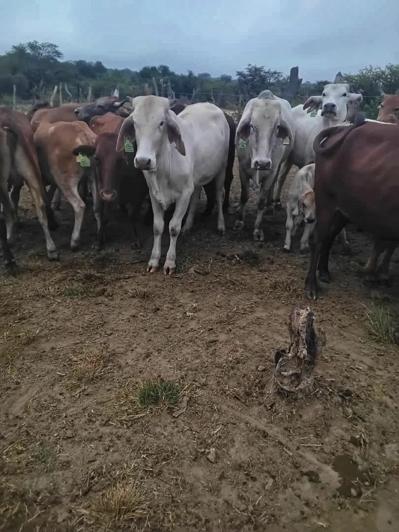
Inspection is the primary method. It can be performed in groups: during rest, feeding, watering, or grazing. During this, sick animals are identified by the fact that they lag behind the herd, feed poorly, show low body condition, appear depressed, and exhibit other symptoms or signs of disease. During an individual inspection, the parts of the animal's body are examined sequentially from head to limbs.
Inspection is best performed in daylight. If necessary, a strong, diffused white light (artificial) can be used. Examination of body contours and their individual parts is performed under side lighting. In some cases, lighting devices (mirror, reflector, endoscope) can be used.
Inspection is performed in a specific sequence: first, the head and neck are examined, then the chest, flank area, abdomen, pelvis, limbs, etc. At the same time, one strives to note any abnormalities in the animal's posture, shape, size, color, and surface properties of the body. When evaluating data obtained by the inspection method, one often resorts to comparing one part of the body with its corresponding opposite.
Inspection can be a very valuable method for mass examination of livestock animals. For example, against the background of a healthy herd, it is easy to notice sick ones, which usually lag behind others and feed poorly.
Palpation (feeling) is an examination method performed by feeling with the fingers. It allows one to obtain an idea of the shape of an organ, its size, consistency, mobility, nature of the surface, sensitivity, pain, humidity, temperature, and other properties. Palpation is used to examine the skin surface and pulse. Palpation is performed with fingertips, the palm, or the fist. Palpation can be:
Palpation provides objective data during a comparative examination of the healthy side with the diseased one. It is best to start palpation from healthy areas and the healthy side, and then move on to the diseased side and the diseased area. Palpation can be performed directly with fingers or with the help of instruments (for example, hoof testers, probes). A distinction is made between deep and superficial palpation.
Superficial palpation is performed by light touches and sliding of the palm over the skin. It can be used to determine the quality of the apex beat, vibration of the chest, surface skin temperature, pain reaction, and muscle tension. It is used for examining pulsating vessels.
Deep palpation involves feeling tissues and organs with the fingertips by applying gradually increasing pressure. This type of palpation is used when examining organs located in the abdominal cavity (stomach, intestines, liver, spleen, and kidneys), especially in small and medium-sized livestock animals.
Varieties of deep palpation include the following methods:
- Penetrating palpation: performed with vertically placed fingers under constant but strong pressure on a limited area (corresponding to the topographical location of the organ being examined).
- Ballottement palpation: consists of short and strong thrusts delivered by the fingers of the hands. It is used to determine fluid in cavities, as well as when examining the liver and spleen. If there is fluid in the abdominal cavity, a fluid thrill is observed, and in the case of diseases of the specified organs, their pain is manifested.
Percussion (tapping) is an examination by delivering rhythmic strikes with a reflex hammer or fingers. The method is based on the different reflection of sound by organs of varying density. A distinction is made between immediate and mediate percussion.
Immediate percussion consists of short strikes on the examined part of the body, delivered by one or several fingers held together and slightly bent. This method is often used to examine the frontal and maxillary sinuses. It is also used in cases where it is necessary to obtain not only auditory but also tactile impressions.
Mediate percussion can be digital or instrumental. Digital percussion is performed with the slightly bent middle finger of the right hand against the terminal phalanx of the index or middle finger of the left hand, which is pressed firmly against the corresponding part of the body (it acts as a pleximeter). The strikes must be short and abrupt. In veterinary practice, digital percussion is applicable when examining small livestock animals, calves, and long-wooled sheep and goats.
Figure 20 – Percussion tools: 1,2 – percussion hammers for large livestock animals; 3,4 – percussion hammers for small livestock animals; 5 – pleximeters.
Instrumental percussion: technique and sound interpretation
Instrumental percussion (tapping) helps to quickly assess the state of an animal's internal organs based on the character of the resulting sound. This method is indispensable for determining organ boundaries and identifying inflammatory foci directly on the farm. For this procedure, a veterinarian needs a pleximeter (a plate made of metal, wood, bone, or plastic) and a percussion hammer. The weight of the hammer is selected according to the dimensions of the patient.
| Type of animal | Hammer weight, g |
| Small animals | 60–75 |
| Large animals | 100–160 |
For accurate diagnostics, it is important to observe the striking technique. The pleximeter is pressed firmly with its entire surface against the animal's body using the left hand. The hammer is held between the thumb and index finger of the right hand so that the handle retains slight mobility. The blow is delivered solely by the movement of the wrist—this allows the hammer to bounce off the plate more easily. The strikes must be short, abrupt, and strictly perpendicular to the pleximeter surface.
The force of the strike is always adjusted to the muscle thickness of the area being examined. For superficial pathological foci in the lungs or when determining the boundaries of internal organs, weak (quiet) percussion is applied. If the animal is in good body condition or if there is a suspicion of deep-seated inflammatory foci, stronger strikes are used. In such cases, it is recommended to use a metal pleximeter.
Tapping is more convenient to perform on a standing animal in a small, enclosed space. During percussion, the examiner's ear should be located strictly at the same height as the pleximeter.
In veterinary practice, two key tapping methods are distinguished. Staccato consists of short, rapid strikes with an instantaneous rebound of the hammer, which allows for the efficient location of inflammatory foci in the lungs. Legato, in contrast, is performed with slow movements with a delay of the hammer on the pleximeter and is used for outlining organ boundaries. Depending on the force of the strike, the depth of physical impact on the tissues also changes.
| Type of percussion | Tissue oscillation depth | Surface oscillation |
| Strong (deep) | up to 7 cm | 4-0 cm |
| Weak | up to 4 cm | 3 cm |
The character of the resulting sound directly indicates the state of internal organs. Organs containing air or gases produce loud and prolonged sounds. For example, a tympanic sound occurs when tapping the cecum in horses and cows or in the area of the paralumbar fossae over the rumen. A clear pulmonary (atympanic) sound is heard over the chest of healthy livestock. Airless tissues — the muscles of the croup or thigh — produce short, quiet, and dull sounds during percussion.
Auscultation: technique of direct and instrumental listening
Auscultation (listening) allows monitoring the function of the heart, lungs, and intestines of animals. With its help, the veterinarian captures natural organ sounds, identifies bronchial rales, friction, or splashing noises in the chest cavity. The functional and morphological state of the organism is judged by the nature of these sounds. In practice, two listening methods are used: direct (with the ear) and indirect (with the help of instruments).
Direct auscultation is performed by applying the ear to the animal's body through a sheet or towel. The front part of a large animal's body on the right side is listened to with the left ear, and on the left side with the right ear. For the veterinarian's safety and the accuracy of the examination, it is important to follow a strict sequence of actions.
- Stand to the side of the animal facing its head.
- Place your hand on the patient's withers or back to control its movements.
- Press your ear firmly against the examined body area.
- When listening to the rear part of the body, turn to face the animal's hindquarters while keeping your hand on its back.
When working near the rear of the animal, use extreme caution to avoid being kicked. For restless horses, the head is restrained and one front leg is raised during the procedure. It is more convenient to listen to sheep, goats, and large dogs on a special table.
Indirect auscultation is performed using stethoscopes and phonendoscopes. Rigid wooden, metal, or plastic stethoscopes conduct sound excellently, especially when examining the heart, but they slide heavily on fur. Flexible stethoscopes with rubber tubes are more convenient for working with any animal, but they can distort and weaken the sound. Phonendoscopes with a membrane that amplifies sounds, as well as combined stethophonendoscopes, are considered the most practical.
Thermometry - measurement of body temperature. Body temperature is determined after rest, before feeding and watering. Thermometer is inserted into the rectum for 5-10 minutes. Elevated body temperature occurs with infectious and parasitic diseases. Lowered temperature occurs in cases of poisoning, milk fever, coma, and in old and emaciated animals.
Table 1 - Limit fluctuations of body temperature in different livestock animal species
Animal species Temperature, °С Animal species Temperature, °С Cattle 37.5-39.5 Rabbit 38.5-39.5 Sheep and goat 38.5-40.0 Fox 38.7-40.5 Pig 38.0-40.0 Mink 39.5-40.5 Horse 37.5-38.5 Arctic fox 39.1-41.4 Donkey 37.5-38.5 Dog 37.5-39.0 Mule 38.0-39.0 Chicken 40.5-42.0 Reindeer 36.0-38.5 Goose 40.0-41.0 Camel 36.0-38.5 Duck 41.0-43.0
Special examination methods: blood pressure determination, electrocardiography, ruminography, radiography, fluorography, stomach probing, catheterization, serological and allergic tests. Laboratory examinations: urine, blood, milk, feces, discharges, secretions, material from deceased livestock animals.
Read next
Veterinary medicine For students
Diagnostics and treatment of rhinitis and laryngitis in livestock animals
Veterinary medicine For students
Fundamentals of veterinary parasitology, classification of zooparasites and types of hosts
Soil and tillage For students