
Etiology, classification and main causes of mastitis in livestock animals
18 min read
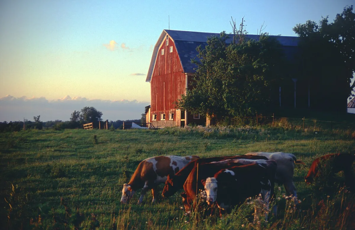
Mastitis is an inflammation of the mammary gland, which most often affects cows, but also occurs in other livestock animals. The disease is not seasonal: it can occur at any stage of lactation, during the dry period, and at any time of the year. The primary damage from mastitis is a sharp decline in milk productivity and the rapid culling of productive animals.
Sources of infection and triggering factors
In most cases, microorganisms are the direct causative agents of the disease or factors that complicate its course. These include streptococci, Escherichia coli, diplococci, Pseudomonas aeruginosa, salmonellae, and mycoplasmas, which act both in isolation and in associations. Infection enters the udder via three main routes:
- galactogenic (through the teat canal) — due to poor hygiene in housing, washing the udder with dirty water or wiping it with a shared towel, pustular lesions on milkers' hands, or a breach of asepsis during catheterization or air inflation;
- lymphogenic (via lymph) — through wounds, abrasions, cracks in the skin of the teats and udder;
- hematogenic (via blood flow) — during gastrointestinal diseases, putrefactive decay of retained placenta, uterine subinvolution, or postpartum endometritis.
Microflora is activated only when the general resistance of the organism and the bactericidal properties of milk are reduced. This is caused by poor feeding, sudden changes in the diet, poor housing conditions, and concomitant diseases. Inflammation is also provoked by mechanical injuries, hypothermia, frostbite, burns, and udder edema.
Errors in milking technology directly lead to mastitis. Excessive vacuum during machine milking, over-milking with teat cups, manual stripping, incomplete milking, irregular milking, and incorrect drying-off are guaranteed to damage tissues and open the path for infection.
Main forms of mastitis and their symptoms
According to their progression, mastitis cases are divided into acute, chronic, and latent (subclinical). The inflammatory process is dynamic: one form can quickly develop into another or take on a mixed character, such as serous-catarrhal, purulent-catarrhal, or purulent-fibrinous. In the absence of treatment, mastitis is complicated by udder induration or gangrene.
In serous mastitis, exudate accumulates in the subcutaneous tissue and interstitial tissue. Usually, 1–2 quarters of the udder are affected: they enlarge, become firm, hot, and painful, and the supramammary lymph node enlarges. Milk initially looks normal, but then becomes watery, and flakes and casein clots appear in it.
Catarrhal mastitis is characterized by lesions of the epithelium of the cistern, milk ducts, or alveoli. In case of cistern catarrh, only the first portions of milk are altered (watery, with casein flakes), while in alveolar catarrh, such milk is produced throughout the entire milking process. Upon palpation, areas of induration are felt in the tissues, but pain is weakly expressed.
Fibrinous mastitis is a severe form of inflammation in which fibrin is deposited in the lumen of the alveoli and ducts. The animal is severely depressed, refuses feed, body temperature rises sharply, and lameness may be noted. The udder is significantly enlarged, becomes hot and very painful, and it is possible to milk only a few drops of yellowish-gray secretion with flakes, films, and traces of blood from the teats.
- Decrease in total milk yield in serous mastitis — 10–30%
- Decrease in milk yield from affected quarters in serous mastitis — 50–70%
- Decrease in milk yield in fibrinous mastitis — 30–80%
- Body temperature in fibrinous mastitis — 40–41 °C
| Form of mastitis | Condition of the animal and udder | Nature of milk and secretion |
|---|---|---|
| Serous | Mild depression, slight rise in temperature. Udder is enlarged, firm, painful; skin is hyperemic. | 10–30% decrease in total milk yield. Milk is watery, with flakes and casein clots (appearing later). |
| Catarrhal | Satisfactory. Areas of induration are felt in the udder tissues; pain is weakly expressed. Teat is doughy. | Milk is liquid, with a bluish or yellowish tint, contains flakes. In alveolar catarrh, flakes appear throughout the milking period. |
| Fibrinous | Depression, refusal of feed, lameness, temperature 40–41 °C. Udder is hot, very painful, crepitus is felt at the base of the teat. | 30–80% decrease in milk yield. Yellowish-gray secretion with fibrin films and blood is released; it is milked with difficulty. |
Specific mastitis includes udder lesions during foot-and-mouth disease, actinomycosis, and tuberculosis. Upon detection of the first signs of inflammation, it is necessary to immediately determine the form of pathology in order to prevent irreversible degeneration of udder tissues.
Purulent-catarrhal mastitis — inflammation of the milk ducts and udder alveoli with the presence of purulent exudate. The acute course of the disease is accompanied by depression of the animal, a sharp decrease in appetite, and an increase in body temperature to 40–41°C.
One, two, or three quarters of the udder are affected; they increase in volume, become painful and hot. Their skin is tense and hyperemic. The supramammary lymph node is enlarged. The total milk yield is reduced by 30-70%.
A small amount of muco-purulent exudate with a thick consistency and white or yellowish flakes is milked from the affected quarter, sometimes a thin, yellow-brown content.
Udder abscesses are characterized by the formation of single or multiple pustules; the latter can be scattered in the tissues of the affected udder quarter or grouped in one or several places; sometimes they merge and dissolve the gland parenchyma.
The general condition of the livestock animal is depressed, appetite is reduced, and body temperature is elevated. The affected udder quarter is swollen, painful, and hot. Abscesses are detected by palpation as follows:
- large abscesses — as hot and painful induration foci, and after their maturation — by fluctuation or fistulous tracts;
- multiple superficially located abscesses — by the uneven surface of the mammary gland;
- small abscesses located deep in the tissues — are poorly palpable.
The supramammary lymph node is enlarged. Milk secretion is reduced by 15-40% overall. If abscesses rupture into the milk ducts, pus and blood are detected in the milk. In the case of superficial abscesses, milk yield decreases, but the quality of the milk by visual signs does not change.
Udder phlegmon is a diffuse purulent or purulent-putrid inflammation of the subcutaneous tissue and interstitial connective tissue of the udder, arising as a complication of serous mastitis, abscesses, and udder injuries. The phlegmonous process develops so rapidly that the udder tissues cannot create a demarcation zone, resulting in the absorption of toxins that cause fever and the general severe clinical picture of the disease characteristic of septic processes.
Usually, half of the udder is affected. It is enlarged, tense, painful, hard, and hot. On the surface of the udder skin, lymphatic vessels protrude in relief as red cords. The supramammary lymph node is significantly enlarged, painful, and has limited mobility.
| Indicator | Value |
| Milk yield | reduced by 50-80% |
| Content (50-100 ml) | watery, sticky, gray color |
Hemorrhagic mastitis is an acute inflammation of the udder characterized by multiple hemorrhages and the infiltration of udder tissues with hemorrhagic exudate. The disease occurs most often in the first days of the postpartum period. The animal exhibits general depression, decreased appetite, and an increase in body temperature. The affected udder quarters are increased in volume, their skin is edematous, covered with red or crimson spots or diffusely hyperemic, and is painful and hot upon palpation. The teat is swollen and edematous. The supramammary lymph node is enlarged and painful upon palpation.
| Indicator | Decrease in milk yield |
| Total milk yield | 25-40% |
| Milk yield from affected quarters | 60-95% |
Milk from diseased quarters is watery, reddish in color ("meat water"), with flakes.
Treatment of cows with mastitis must begin no later than the first 24 hours from the appearance of the first signs of inflammation. The following complex of measures is applied:
- The sick animal is isolated and placed in a clean, bright, warm stall with dry, soft bedding.
- Succulent feed is excluded from the diet, replaced with good hay, and the amount of concentrated feed is reduced to the minimum daily allowance; water intake is restricted.
- The cow is switched to manual milking, performed every 6-8 hours.
- In the presence of concomitant diseases (uterine subinvolution, metritis, feed poisoning, etc.), measures are taken to eliminate them.
In the initial stages of mastitis, one of the methods of novocaine therapy is used: nerve blockade of the udder according to D.D. Logvinov or B.A. Bashkirov, suprapleural blockade according to V.V. Mosin, or intra-aortic administration of a 1% novocaine solution (100 ml) according to D.D. Logvinov and N.D. Volvach.
Udder nerve blockade in cows according to D.D. Logvinov is carried out by injecting a 0.5% novocaine solution into the connective tissue space between the abdominal wall and the base of the affected udder quarter. If the front quarter is inflamed, the needle is inserted from the anterolateral surface of the udder in a place where a groove is easily felt at the border between the base of the gland and the abdominal wall. The injection site is pre-clipped and wiped with a swab moistened with alcohol or smeared with a 5% iodine solution. The needle is advanced into the supramammary space to a depth of 8-10 cm parallel to the abdominal wall towards the knee joint of the opposite side. The needle is moved in different directions, and 100-200 ml of sterile 0.5% novocaine solution is injected (using a Janet syringe connected to the needle via a rubber tube).
In case of inflammation of the hind quarter, the needle is inserted from the rear surface of the udder above its base, retreating 2-3 cm from the midline towards the affected quarter. The needle is directed towards the carpal joint of the same side. Otherwise, the procedure is the same as for the front quarter blockade.
To quickly relieve pain and suppress inflammation in mastitis, a suprapleural novocaine blockade of the udder is used. The method is based on injecting an anesthetic solution into the space between the major and minor psoas muscles. In this case, the external spermatic nerve, which serves as the main source of udder innervation, and branches of the ilioinguinal nerve are simultaneously blocked. If both halves of the organ are affected, a bilateral blockade is performed.
Before starting the procedure, the cow must be securely restrained by the horns or using nose tongs. The injection point is determined in the space between the transverse processes of the 3rd and 4th lumbar vertebrae, 7–8 cm away from the midline of the body. In animals with moderate body condition, the outer edge of the longissimus dorsi muscle is clearly visible in this area. The skin in the injection zone is prepared according to standard aseptic rules.
- With a sharp movement, insert the needle perpendicular to the skin at the marked point.
- Advance the needle 6–8 cm deep at an angle of 55–60° to the median plane of the body until it rests against the vertebral body.
- Attach a syringe with the novocaine solution to the needle and, while applying slight pressure to the plunger, pull the needle back 25 mm.
- Slowly inject 80–100 ml of the solution.
The main sign that the needle is exactly in the intermuscular connective tissue space is the free movement of the syringe plunger during the administration of the drug.
Anesthesia of the corresponding half of the udder occurs within 10–15 minutes and lasts for 2–3 hours. It should be noted that pain sensitivity partially remains in the area of the milk mirror and at the base of the rear teats. To remove stasis, the novocaine block can be combined with physiotherapy: applications of heated sapropel, dry heat in the form of a thiosulfate heating pad, ultrasound therapy, or galvanic ion therapy.
Surgical treatment of complications and general drug therapy
- Novocaine concentration for block — 0.5%
- Single dose of novocaine — 80–100 ml
- Duration of anesthesia — 2–3 hours
- Course of antibiotic treatment — 4–5 days
- Interval for calcium chloride administration — 12 hours
When inflammation transitions into a purulent form with the formation of abscesses, surgical intervention is required. Superficial abscesses are opened with vertical incisions. After removing the contents, the wound is dusted with streptocide powder or a gauze drain soaked in turpentine is inserted for 1–2 days, changing it 2–3 times a day. Subsequently, the cavity is washed with antiseptic solutions and lubricated with antimicrobial emulsions or ointments.
From deep abscesses, pus is extracted with a syringe and needle. Through the same needle, the cavity is washed with solutions of penicillin, neomycin, bicillin, furacilin, or ethacridine lactate. To accelerate encapsulation and neutralize the inflammatory focus, 20–50 ml of a 5% alcoholic iodine solution is injected into the abscess cavity after washing.
If mastitis progresses with signs of severe intoxication and a risk of sepsis, the animal is prescribed intravenous administration of a complex mixture. The solution is prepared immediately before administration and is used 1 to 3 times throughout the treatment course.
| Component of the mixture | Drug concentration | Volume per injection |
|---|---|---|
| Glucose | 40% | 400 ml |
| Urotropine | 40% | 30 ml |
| Caffeine | 20% | 10 ml |
| Calcium chloride | 10% | 120 ml |
In cases of serous and catarrhal mastitis, local cooling of the affected quarter using compresses or applications of liquid clay is indicated in the very first hours of the disease. If udder edema progresses, a 10% calcium chloride solution is administered intravenously at a dose of 150–200 ml every 12 hours to reduce capillary permeability. For the serous form, a single drenching of sodium sulfate at a dose of 400–800 g is also effective.
To combat systemic infection during high body temperature, a course of intramuscular antibiotic injections is mandatory, calculated at 3–5 thousand IU per 1 kg of the animal's body weight. The effectiveness of the therapy depends on the correct choice of active ingredient. Treatment is continued for 4–5 days.
Before starting antibiotic use, be sure to submit milk from the affected udder quarter for bacterial culture to determine the sensitivity of the microflora to specific drugs.
In severe cases, especially with purulent mastitis, intramuscular antibiotic injections are combined with intravenous administration of sulfonamides. For this, a 10% sodium norsulfazole solution is used at a dose of 120–150 ml every 12–24 hours. A total of 3–4 such injections are prescribed for the course.
Local therapy and physiotherapeutic methods for mastitis
When flakes and clots are detected in the milk, which is characteristic of catarrhal, purulent-catarrhal, fibrinous, and hemorrhagic mastitis, local treatment must be performed. The main method of fighting pathogens in the affected udder quarter is the intracisternal administration of antimicrobial and anti-inflammatory drugs. For the procedure to be successful, it is important to strictly follow the technology of udder preparation and drug dosage.
- Heating temperature of drugs before administration — 37–40 °С
- Milk withdrawal period after antibiotics — 3–5 days
- Dose of oxytocin for emptying the udder quarter — 30–60 IU
- Timeframe for latent mastitis test after treatment — 7–10 days
- Prepare the udder quarter. Thoroughly milk out the quarter. If flakes and mucus hinder milking, inject 100 ml of saline-soda solution into the teat cistern (1 g of sodium chloride and 2 g of baking soda per 100 ml of distilled water; 1 g of trypsin or pepsin can be added additionally). Perform a light massage and milk out the secretion after 15–20 minutes. For maximum emptying of the diseased quarter, inject the cow with 30–60 IU of oxytocin subcutaneously 5–7 minutes before milking or intravenously immediately before it.
- Disinfect the teat. Wipe the tip of the teat with a swab soaked in 70% alcohol.
- Administer the drug. Heat the drug in a water bath to a temperature of 37–40 °С. Introduce the agent intracisternally using a sterile shortened milk catheter.
- Perform a massage. After each administration, gently massage the teat and udder in an upward direction for better distribution of the drug in the tissues.
During antibiotic treatment and for 3–5 days after its completion, milk obtained from the cow must not be used for food.
| Drug / Active ingredient | Dosage | Application scheme |
|---|---|---|
| Antibiotic solutions in a 0.25–0.5% novocaine solution or isotonic sodium chloride solution | 100–150 ml | Intracisternally, 1–3 times a day, course 2–4 days |
| Mastisan-A, -B, -E | 10–15 ml | Intracisternally, 1–3 times a day, course 2–4 days |
| Masticid | 15–20 ml | Intracisternally, 1–3 times a day, course 2–4 days |
| 1% solution of soluble streptocide | 100–150 ml | Intracisternally, 1–3 times a day, course 2–4 days |
| 2–3% solution of norsulfazole sodium | 100–150 ml | Intracisternally, 1–3 times a day, course 2–4 days |
| Furacilin solution (1:5000) | 100–150 ml | Intracisternally, 1–3 times a day, course 2–4 days |
| 2–4% propolis ointment on an oil-lanolin base or PEG-400 | 5–7 ml | Intracisternally, 1–3 times a day, course 2–4 days |
| Penicillin or erythromycin (for subclinical mastitis) | 50–100 thousand units | Intracisternally, three times with an interval of 24 hours |
| Mastisan-E or other antimicrobial drugs (for subclinical mastitis) | According to manufacturer's instructions | Intracisternally, three times with an interval of 24 hours |
Udder massage is an important part of therapy, but its technique strictly depends on the nature of the inflammation. In case of serous mastitis, massage is performed from the bottom up — from the tip of the teat to the base of the udder, stimulating lymph drainage. In case of catarrhal mastitis, the direction is reversed — from top to bottom, aiding the removal of flakes. If fibrous mastitis is diagnosed in the stage of infiltrate resorption (in the absence of pain), a mixed technique is used in both directions. Remember that massage is completely contraindicated for abscesses, phlegmon, udder gangrene, and also in the initial stages of purulent-catarrhal, hemorrhagic, and fibrinous processes.
With subclinical mastitis, there are no obvious signs of inflammation, but milk yield decreases and milk quality deteriorates. The subclinical form is diagnosed by changes in the physicochemical properties of the raw material and by the results of bacteriological examination to determine the quantity and type of microbes per 1 ml. Treatment is carried out by administering antimicrobial agents, combined with physiotherapy. Follow-up testing is scheduled 7–10 days after the course of treatment, and in case of a positive result, therapy is repeated until full recovery.
The following physiotherapeutic methods are suitable for treating subclinical mastitis:
- applications of heated ozokerite or paraffin to the affected quarter;
- warming bandages;
- heating with Solux or Infrareux lamps.
Complications, chronic course of the disease, and mastitis in mares
If acute mastitis is not treated systematically, the disease progresses to a subacute and then to a chronic form within 5–7 days. The most common is chronic purulent-catarrhal mastitis, which often develops covertly, without a pronounced acute stage. The general condition of the cow remains satisfactory, the local temperature is not elevated, and tenderness upon palpation is weakly expressed. Slow degeneration of the parenchyma occurs in the udder tissues, with its replacement by connective tissue, causing the quarter to decrease in volume, become firm, and milk yield to progressively drop.
During the chronic purulent-catarrhal process, the secretion acquires a distinct muco-purulent character. Treatment of this form is ineffective and requires considerable time. Veterinary specialists may use intracisternal antibiotics in conjunction with thermal applications (ozokerite, paraffin), ultrasound, or iontophoresis. However, due to low economic efficiency, cows with chronic mastitis are most often culled.
The situation is similar with udder induration — a severe complication of mastitis accompanied by the proliferation of connective tissue. The affected quarter becomes stony, painless, and does not decrease in size even after milking or during the dry period. Since treatment for induration does not yield results, and the milk becomes watery and unpleasant in taste, such animals are also culled.
Mastitis in mares has its own specifics and occurs mainly on kumis farms, proceeding primarily in a catarrhal or purulent-catarrhal form. The affected part of the udder swells, enlarges, becomes hot, painful, and acquires a doughy or firm consistency. Instead of normal milk, a watery fluid with casein flakes or a muco-purulent mass is secreted. Affected mares refuse feed, experience fever, limp when moving, and do not allow foals to suckle, standing with their hind limbs spread apart.
When the process progresses to a purulent form in mares, multiple or solitary abscesses form rapidly. The cavities of these abscesses fill with purulent exudate and necrotic udder tissues. Over time, the abscesses mature and rupture, forming deep fistulas.
When examining affected mares, first rule out udder botryomycosis. This chronic disease is accompanied by the proliferation of connective tissue, causing the affected half or the entire udder to turn into a dense tumor with ulcers and deep fistulous tracts. Unlike common purulent mastitis, botryomycosis involves the formation of multiple painless nodules that exude a gray, staining exudate. The diagnosis is confirmed by microscopy of the pus for the presence of fungal colonies.
In mares, each teat has 2 to 3 teat canals. For local treatment, drugs must be administered through each teat opening using a thin milk catheter.
- Isolate the foal from the mare and switch it to artificial feeding with milk from other mares or healthy cows.
- Ensure frequent milking of the affected half of the udder.
- Administer warmed antibiotic solutions in novocaine or ready-made suspensions through the teat canals.
- Perform a supra-mammary novocaine block and begin a course of intramuscular antibiotic injections.
- Apply heat to the udder (paraffin, ozokerite) and open matured abscesses.
In cases of udder botryomycosis and the development of multiple abscesses, treatment of mares is most often ineffective.
Therapy regimens for mastitis in sows, sheep, goats, and rabbits
In pigs, inflammation usually affects individual segments, half, or the entire gland, occurring in serous, serous-catarrhal, or purulent forms. The affected areas increase in volume and become red, hot, painful, and firm, while the milk becomes watery with traces of flakes or pus. In cases of purulent mastitis, the sow is depressed, has a fever, refuses feed, and does not allow piglets to suckle.
In sheep and goats, the same forms of mastitis are recorded as in cows. When serous, catarrhal, or purulent-catarrhal inflammation develops, a short novocaine block with an antibiotic shows high efficiency. If a gangrenous process has started in the udder, it is almost impossible to stop it conservatively.
In rabbits, purulent inflammation with the formation of abscesses that turn into ulcers develops more often. Udder phlegmon is extremely severe: the skin turns blue, black, and hard to the touch. With phlegmon, animals most often die, and upon recovery, extensive scars remain in the tissues.
In cases of udder gangrene in sheep and goats, as well as phlegmon or damage to more than 2–3 gland segments in rabbits, the prognosis is unfavorable. Such rabbits are culled, and small ruminants are operated on (udder amputation or removal of necrotic tissues).
- Novocaine concentration for blocks — 0.5%
- Single dose of Bicillin-3 for sheep — 600,000 units
- Dose of antibiotics for rabbits — 15,000–20,000 units/kg
- Interval between blocks in mares — 48 h
| Animal species | Novocaine blocks | Drugs and dosages | Supportive measures |
|---|---|---|---|
| Mares | Supra-mammary block: 100–150 ml of 0.5% novocaine solution above the base of the affected half (injection from the front along the abdominal wall). Repeat after 48 hours. | Intramuscularly: antibiotics. Into teat canals: penicillin with streptomycin or neomycin with tetracycline, 100,000–200,000 units in 20–30 ml of 0.5% novocaine solution 1–2 times daily, or Mastisan, Masticid. |
Frequent milking, heat (ozokerite, paraffin) on the udder, opening of matured abscesses. |
| Pigs | Short block: 30–50 ml of 0.5% novocaine solution above the base of each affected segment. | Intramuscularly: antibiotics. Orally: calomel, hexamethylenetetramine, phenyl salicylate. Topically: rubbing with camphor or ichthyol ointment. |
Frequent milking of affected segments, heat (heated ozokerite, paraffin), opening of matured abscesses under a glue bandage. |
| Sheep and goats | Short block: 40–60 ml of 0.5% novocaine solution with the addition of 600,000 units of Bicillin-3. Repeat after 3–4 days if necessary. | Intramuscularly: antibiotics. Intravenously (for gangrene): Kadykov's fluid. |
Therapy is similar to the treatment of cows. Surgical removal of necrotic tissues or udder amputation for gangrene. |
| Rabbits | Short block: 3–5 ml of 0.25–0.5% novocaine solution under the base of each diseased segment. | Intramuscularly: penicillin and streptomycin, 15,000–20,000 units per 1 kg of body weight 2–3 times daily. | Isolation from kits, milking of secretion (at the beginning of the disease before abscesses), opening of abscesses with irrigation using disinfectant solutions and dusting with streptocide. |
Read next
Veterinary medicine For students
Diagnostics and treatment of rhinitis and laryngitis in livestock animals
Livestock For students
Methods of accounting and quality control of milk in dairy cattle breeding
Veterinary medicine For students