
Anatomical structure of fruit tree branches and tissue functions
2 min read
Any mistake when working with secateurs or a grafting knife can damage the conducting pathways and destroy the shoot. To avoid this, an agronomist must be precisely oriented in the structure of the branches. Understanding the anatomy of the cut allows one to correctly assess the viability of a branch and perform pruning or grafting with minimal trauma to the plant.
The degree of winter damage to branches can be determined by the color of the tissues on a cross-section. Healthy tissues remain light, while areas of wood damaged by frost darken due to the destruction of conducting vessels.
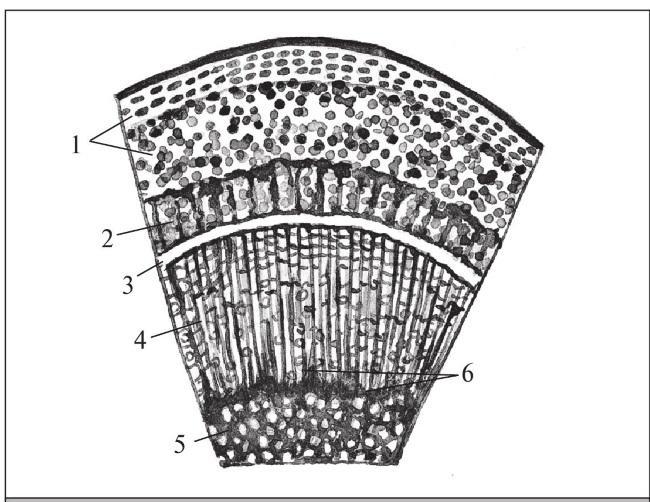
Anatomical layers of a branch and their functions
In the very center of the branch is the pith, consisting of uniformly developed parenchymal cells and intercellular spaces. By autumn, the pith cells fill with stored nutrients. This reserve is vital for successful overwintering and the spring start of shoot growth.
The pith is surrounded by multi-layered wood (xylem). Through its vessels and tracheids flows the upward transport of water and mineral salts from the roots to the leaves and fruits. The strength of the trunk and branches is provided by the thick-walled mechanical cells of the wood, while the wood parenchyma and pith rays accumulate nutrients by autumn.
The layer of wood is surrounded by a thin ring of cambium. The cells of this tissue possess a high ability to divide: they deposit wood cells inward and phloem cells outward. The cambium is the most active tissue of the tree, ensuring its regeneration. Due to its activity, a shoot is capable of recovering even if the wood is completely destroyed, through the growth of new conducting layers.
On the outer side of the cambium is the bast (phloem). Through its conducting elements, a downward flow occurs — the outflow of plastic substances from the leaves to other organs of the above-ground and root systems. This conducting tissue consists of three main elements:
- parenchymal cells;
- sieve tubes with companion cells;
- fibers of mechanical tissue — bast fibers.
The upper protective layer of the branch is represented by the bark, which consists of cork tissues and parenchymal cells. Cork protects the inner layers of the tree from freezing, pests, and mechanical damage. The parenchymal cells of the bark serve to accumulate stored nutrients for the winter period.
Read next
Pruning and training For students
Determining the age of fruit trees and individual branches by external features and cross-sections
Pruning and training For students
Anatomy and physiology of the leaf apparatus of fruit trees during canopy formation
Pruning and training For agronomists