
Biochemical composition and quality indicators of vegetable oils
18 min read
Accumulation of vegetable oils and their role in crop survival
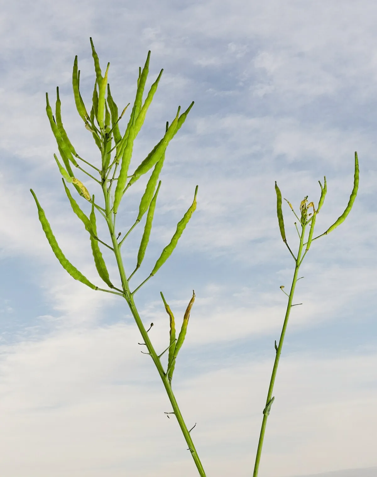
Vegetable fats and oils are esters of the trihydric alcohol glycerol and higher carboxylic acids (triacylglycerols). The difference between them is physical: fats remain solid at room temperature, while oils retain a liquid consistency due to the predominance of unsaturated fatty acids. In the vegetative organs of plants, the concentration of fats rarely exceeds 5% dry matter, whereas in the seeds of oil crops it can be higher than 50%.
The oxidation of fats releases more metabolic water than the breakdown of proteins or carbohydrates. This internal resource increases plant survival under water stress conditions.
Vegetable oils consist of mixtures of triglycerides, which can be monoacid or polyacid. Monoacid glycerides contain residues of only one acid (for example, oleic, ricinoleic, or stearic), but they are extremely rare in nature. The vast majority of oils in practice are mixtures of polyacid glycerides with a small amount of accompanying substances.
- Glycerides in crude oil — 95–98%
- Free fatty acids — 1–2%
- Phosphatides — up to 1–2%
- Sterols — 0.3–0.5%
| Crop | Oil content, % of dry matter | Crop | Oil content, % of dry matter |
|---|---|---|---|
| Sunflower | 29–56 | Sesame | 48–63 |
| Safflower | 25–37 | Perilla | 26–49 |
| Indian mustard | 35–47 | Lallemantia | 29–37 |
| Winter rapeseed | 45–49 | Peanut | 41–55 |
| Camelina | 25–46 | Soybean | 15–24 |
| Castor bean | 47–58 | Oil flax | 30–49 |
Technological quality indicators and oil processing
The properties of vegetable fat are determined by its melting point, acid value, iodine value, and saponification value. The melting point depends directly on the acid composition: the prevalence of saturated acids (palmitic, stearic, myristic) makes the fat solid, while the prevalence of unsaturated ones (linoleic, linolenic) makes it liquid. The acid value indicates the amount of free fatty acids that were not used in biosynthesis during seed ripening or were formed during oil spoilage.
The acid value is a critical indicator of harvest and oil shelf life, as it easily increases during storage. For top-grade sunflower oil, this indicator should not exceed 1.3 mg KOH, for the first grade — 2.2 mg KOH, and for the second grade — 5 mg KOH.
The iodine value reflects the degree of unsaturation of fatty acids and shows how many grams of iodine 100 grams of fat can bind. In vegetable oils, this indicator is high, ranging from 120–160, whereas in animal fats it fluctuates between 30–70. Based on the iodine value, vegetable oils are divided into two groups:
- drying (iodine value ≥130) — for example, flaxseed and hemp;
- non-drying (iodine value <85) — castor and peanut.
The higher the iodine value, the more liquid the oil is, the easier it oxidizes, and the more suitable it is for the production of drying oil, varnishes, and paints. The oil also contains carotenoids and vitamins.
In the food industry, liquid vegetable oils are converted into solid fats for margarine production through hydrogenation. In this process, hydrogen, in the presence of catalysts, adds to the double bonds of unsaturated acids, converting them into saturated ones. Another important reaction is saponification under the action of acids or alkalis, resulting in the formation of free glycerol and free fatty acids or their salts. The average molecular weight of acylglycerols in fat is estimated by the saponification value — the number of milligrams of potassium hydroxide required to neutralize all free and bound acids in 1 gram of oil.
Why vegetable oils spoil during storage
During long-term storage, vegetable fats inevitably spoil, acquiring a bitter taste and a specific odor. This process is caused by both chemical reactions under the influence of the external environment and enzymes. For example, under the influence of the enzyme lipase, fat hydrolysis occurs, analogous to the saponification reaction. This releases free fatty acids, which impair the taste of the oil, and its acid value increases.
The most common cause of spoilage is the oxidation of unsaturated fatty acids by atmospheric oxygen. Oxygen adds to the double bonds, forming peroxides. In the course of their further decomposition, aldehydes are released, which give the oil an unpleasant odor and a rancid taste.
The process of fat oxidation accelerates sharply in the presence of humidity, with an increase in temperature, and under the influence of light. To prevent rancidity, oil is stored without access to air (in a vacuum) or antioxidants such as tocopherol (vitamin E) are added to it.
Plant protective lipids: waxes, cutin, and steroids
The outer surface of leaves, stems, and fruits is protected by a cuticle — a waterproof barrier made of cutin and wax. The wax bloom on grapes, plums, pears, and apples protects the tissues from drying out, excessive wetting, as well as from the penetration of pathogens and pests. Waxes are synthesized in the cytoplasm of epidermal cells, from where they are secreted to the outside in the form of microscopic plates, granules, or rods.
From a chemical point of view, waxes are esters of higher monohydric alcohols and higher fatty acids. They are the most hydrophobic lipids in a plant and can contain up to 50% impurities. Their composition includes free higher alcohols with an even number of carbon atoms (from C22 to C32), free long-chain higher fatty acids (from C24 to C36), saturated hydrocarbons with an odd number of carbon atoms (from C21 to C37), as well as pigments and aromatic substances.
| Characteristic wax fatty acids | Chemical formula |
|---|---|
| Carnaubic | C24H48O2 |
| Montanic | C29H58O2 |
| Cerotic | C27H54O2 |
Among the high-molecular alcohols included in the structure of plant waxes, the following compounds predominate:
- cetyl alcohol — CH3(CH2)14CH2OH;
- myricyl alcohol — C31H63OH;
- n-hexacosanol — CH3(CH2)24CH2OH;
- n-octacosanol — CH3(CH2)26CH2OH;
- n-triacontanol — CH3(CH2)28CH2OH.
- Wax in sunflower seed coat — 0.2%
- Wax in flax seed coat — 0.03%
- Wax in soybean seed coat — about 0.01%
Although the seeds of oil crops contain little wax, the leaves of some plants excrete it in such quantities that the raw material is harvested for industrial candle production.
Cutin and suberin serve as an insoluble framework in which waxes are embedded. Cutin forms a multi-layered cuticle on epidermal cells, consisting of epicuticular wax, the cuticle proper, cuticular layers (a mixture of cellulose, cutin, and wax), and a pectin layer. Suberin strengthens the cell walls of the cork, Casparian strips in the endodermis, and vascular bundles in grasses, forming a layered structure with water-repellent properties.
Steroids belong to terpenoids and act as regulators of biochemical processes. Their structure is characterized by the presence of an oxygen-containing substituent at C3, methyl groups at C10 and C13, as well as the absence of double bonds in the rings. This group includes sterols, steryl esters, sapogenins, hormones, and ergosterol (vitamin D). Most steroids are present in cells in trace amounts; however, the concentration of sterols can in some cases exceed 2%.
Steryl esters and pigments: the framework of photosynthesis and protection against overheating
The functioning of the plant photosynthetic apparatus and the strength of cellular structures are largely determined by lipophilic compounds — sterols and steryl esters. Sterols, which are present in corn leaves and wheat germ, contain 8 to 10 carbon atoms in the side chain at C17 and a hydroxyl group at the C3 position. Inside the cell, they form complex structures with proteins, creating the basis of intracellular membranes. Steryl esters are esters of these sterols and higher fatty acids (palmitic, stearic, oleic). They are also bound to proteins, insoluble in water, but dissolve perfectly in fat solvents, ensuring the stability of the cell framework.
Key plant pigments — chlorophylls and carotenoids — are also concentrated in membrane structures. To date, about 10 types of chlorophylls are known, differing in structure and color. Chlorophylls a (blue-green) and b (yellow-green) are always present in the cells of higher green plants. By comparison, diatoms contain chlorophyll c, red algae contain chlorophyll d, and photosynthetic bacteria utilize four bacteriochlorophylls (a, b, c, and d).
The chlorophyll molecule is held within the chloroplast due to its dual nature: its polar hydrophilic porphyrin "head" faces the proteins, while the hydrophobic phytol "tail" is anchored in the lipid layer.
Chemically, chlorophyll is structured as an ester of the dicarboxylic organic acid chlorophyllin and two alcohols — methyl (CH3OH) and phytol (C20H39OH). Chlorophyllin itself belongs to the magnesium porphyrins and is described by the empirical formula MgN4OH30C32(COOH)2. The formula for chlorophyll a is C55H72O5N4Mg, and for chlorophyll b is C55H70O6N4Mg. Due to the replacement of one methyl group with an aldehyde group, chlorophyll b contains two fewer hydrogen atoms and one more oxygen atom.
Next to chlorophyll in the lipid layer of the membranes are carotenoids — accessory pigments that protect the plant from light-induced damage. They all consist of 40 carbon atoms and are derivatives of isoprene (CH2=C(CH3)–CH=CH2). Thanks to the system of conjugated bonds, these polyene pigments have red, orange, or yellow colors and are characterized by high chemical activity. Carotenoids are divided into carotenes with the general composition C40H56 and oxygen-containing xanthophylls C40H56O2. By absorbing short-wave rays, they act as a shield against destructive vitacidal radiation.
- Chlorophyll a to b ratio — 3 times higher
- Number of carbon atoms in carotenoids — 40
- Share of phosphatidylglycerol in leaf lipids — up to 50%
Phospholipids: the basis of cell membranes and stress resistance
Phospholipids (phosphatides) determine the permeability of cell membranes and the ability of plants to withstand drought and low temperatures. They differ from ordinary fats by the presence of a phosphoric acid residue and a nitrogen-containing fragment. They contain residues of fatty acids, such as linoleic, linolenic, palmitic, and stearic acids. Based on the nature of the polar group (component X), phospholipids are divided into several groups.
- Choline phospholipids (lecithins) — contain choline, which is soluble in water and alcohol and capable of transferring methyl groups during metabolic processes.
- Colamine phospholipids (cephalins) — contain colamine.
- Serine phospholipids — contain the amino acid serine.
- Inositol phospholipids — contain the alcohol inositol.
A special group consists of phosphatidic acids, which lack nitrogenous bases. They are found in cabbage leaves, wheat germ, and the latex of rubber-bearing trees in the form of potassium, magnesium, and calcium salts. By attaching another glycerol residue, they are converted into phosphatidylglycerol. This substance accumulates in the chloroplasts of leaves and plays a vital role in their vital functions.
Since phospholipids have polar hydrophilic heads and insoluble hydrophobic tails, they self-organize into a double layer in the aqueous environment of the cell. The breach of this membrane barrier under unfavorable conditions leads to the loss of water and nutrients by the cell.
Layered structures formed by phospholipids in solution play an important role in the construction of cell membranes. Phospholipids are natural antioxidants; they protect oils from oxidation by easily oxidizing themselves. The content of phospholipids in plants is insignificant. The highest amount is found in soybean seed – 1.5–2.0%.
Glycolipids. Glycolipids contain either glycerol or sphingosine as the alcohol component, as well as a carbohydrate component and fatty acid residues. Unlike phospholipids, they do not contain a phosphoric acid residue. The carbohydrate component in glycolipids can be glucose, galactose, glucosamine, galactosamine, and their acetyl derivatives, or oligosaccharide chains consisting of the listed monosaccharides. The higher fatty acids that make up glycolipids are quite diverse. The most important representatives of the glycolipid class are monogalactosyl diacylglycerols, digalactosyl diacylglycerols, and trigalactosyl diacylglycerols. The first two glycolipids were initially isolated from wheat, and the third from potato tubers. The bulk of cellular glycolipids are located in chloroplasts, with only insignificant amounts present in mitochondria. Glycolipids in chloroplasts are localized in the membranes; the ratio of monogalactosyl diacylglycerols to digalactosyl diacylglycerols in the chloroplast envelope membranes is approximately 0.9:1, whereas in the membranes of the thylakoid system, it is typically 2:1.
Sulfolipids. In terms of structure, sulfolipids are close to glycolipids. The only representative of this class present in plant tissues is the sulfonolipid, often called a sulfolipid. In the molecule of this compound, the glycosyl residue 6-sulfo-6-deoxy-D-glucose is connected by an α-glycosidic bond to the sn-3 position of 1,2-diacyl-sn-glycerol. It should be noted that the glycosyl residue is a sulfonate (C–S bond) rather than a sulfate ester (C–O–S bond). In this compound, the predominant fatty acid is α-linoleic C18:2, but palmitic acid C16:0 may also be present. In plants, the sulfonolipid is localized in chloroplasts, primarily in the membranes of the thylakoid system. In smaller quantities, it has been detected in etioplasts and proplastids. Very small amounts of this lipid (about 1% of their total quantity) are contained in potato tubers and apple fruits.
Cerebrosides. Cerebrosides consist of monosaccharide-galactose residues attached by a glycosidic bond to the first carbon atom of an N-acylated derivative of sphinganine–ceramide. Cerebrosides have been detected in wheat grain, bean leaves, potato tubers, apple fruits, and fungi.
Organic solvents are used for the extraction and separation of lipids; they not only dissolve lipids but are also capable of breaking down lipoprotein complexes, thereby achieving the extraction of both free lipids and lipids that are part of these complexes. The degree of lipid recovery can reach 96–98%. Petroleum ether, diethyl ether, and chloroform are most often used for lipid extraction. Mixtures of solvents of varying polarity are also used, for example, chloroform–methanol, ethanol–diethyl ether, etc. As a rule, the use of solvent mixtures leads to a more complete extraction of lipids from various tissues.
Various chromatographic methods are used for the separation of neutral lipids. Silicic acid and silica gel are most often used for the separation of neutral lipids. By impregnating a thin layer of silica gel with a silver nitrate solution, it is possible to increase the resolving power of the adsorbent and achieve the separation of glycerides by their degree of unsaturation.
Phospholipids are separated using their different solubility in organic solvents. The isolation of "lecithin" and "cephalin" fractions is possible precisely due to this property. These fractions are not pure but represent mixtures of phospholipids with similar physicochemical properties, enriched with phosphatidylcholines (the former) and phosphatidylethanolamines (the latter). Usually, phospholipids are separated by adsorption chromatography on silicic acid or silica gel (in a thin layer or columns); DEAE- and TEAE-cellulose are also used. During column chromatography on silicic acid (with an increasing proportion of methanol in the eluent composition), phospholipids are separated in the following sequence: phosphatidylethanolamines, phosphatidylinositides, phosphatidylcholines, sphingomyelins. The obtained fractions can be further purified by TLC; however, even after such purification, each spot on the chromatogram contains phospholipids that differ in chain length and the degree of unsaturation of fatty acid residues, as well as in the types of bonds of hydrophobic components with the polyol (diacyl, plasmalogen, or alkyl phospholipids).
As with neutral lipids, an effective method for separating phospholipids by the degree of unsaturation of fatty acid residues is chromatography in adsorbent layers impregnated with a silver nitrate solution.
Methods for determining lipid content are based on their ability to dissolve in various organic solvents. Free lipids are extracted from the analyzed material with nonpolar solvents (hexane, diethyl ether), while bound lipids are extracted with solvent systems usually containing alcohol (a mixture of chloroform and methanol in a 2:1 volume ratio). Tightly bound lipids are obtained from the meal treated with alkalis and acids that remains after the extraction of bound lipids.
The most convenient method for determining the crude fat content in seeds and vegetative plant organs is the method proposed by Soxhlet. Fat extraction is performed with ethyl (sulfuric) ether. The extracted crude fat is freed from the solvent and weighed. The composition and quality of fats and oils are characterized using various analytical "numbers," implying the consumption of specific reagents for reactions with the fat. The most significant are the following numbers: acid, saponification, iodine, and peroxide.
Chemical quality control: four main indicators of vegetable oil
During the storage of oilseed crops and finished vegetable oil, latent chemical processes occur. Due to the influence of humidity, heat, and air, fats gradually hydrolyze and oxidize, which reduces product quality. To timely detect these changes and determine the direction of processing, laboratories monitor four main indicators.
- Acid value — a measure of free acid accumulation
- Saponification value — an indicator of the total acid concentration
- Iodine value — a measure of unsaturation and drying capacity
- Peroxide value — an indicator of the initial stage of spoilage
Acid value is a key indicator of oil and seed quality. It increases during long-term storage of raw materials due to fat hydrolysis. To determine this value, free fatty acids in the oil are neutralized with a titrated potassium hydroxide solution. A reaction occurs between the alkali and the acids: RCOOH + KOH → RCOOK + H2O, where RCOOH is a fatty acid. The acid value is judged by the amount of KOH solution consumed for neutralization.
An increase in the acid value indicates a violation of seed storage conditions. If measures are not taken in time, self-heating of the bulk and deep decomposition of triacylglycerols will begin.
Saponification value is determined to establish the type of fat. During the analysis, a sample is boiled with an excess of titrated potassium hydroxide solution, resulting in fat hydrolysis. The released fatty acids immediately react with potassium hydroxide. The excess alkali that did not react with the acids is titrated with hydrochloric acid: KOH + HCl → KCl + H2O. The saponification value is calculated based on the amount of alkali consumed to bind all the fat acids.
Iodine value is necessary to determine the type of fat and its ability to dry. This indicator is also used to calculate hydrogen consumption for oil hydrogenation. Most often, the iodine value is determined by the bromometric method:
- A solution of bromine in anhydrous methyl alcohol saturated with sodium bromide is used.
- Bromine forms an unstable complex compound with sodium bromide: NaBr + Br2 → NaBr · Br2.
- By detaching from the complex, bromine reacts with unsaturated glycerides.
- The amount of unreacted bromine is determined iodometrically and the iodine value is calculated.
Peroxide value serves as the main indicator of oxidative spoilage of fats. In the presence of atmospheric oxygen, the acids that make up the oil are partially oxidized to form peroxides. This process is also observed during oil drying. The indicator is expressed in grams of iodine that can react with the peroxides contained in 100 g of fat. The determination is based on the fact that fatty acid peroxides react with potassium iodide, releasing free iodine, which is then titrated with a hyposulfite solution.
Read next
Agrochemistry For students
The effect of mineral nutrition on fat accumulation in crops
Agrochemistry For students
Chemical structure and classification of fatty acids in plants
Agrochemistry For students